4ZYN

Crystal Structure of Parkin E3 ubiquitin ligase (linker deletion; delta 86-130)


Experimental Data Snapshot

  • Method: X-RAY DIFFRACTION
  • Resolution: 2.54 Å
  • R-Value Free: 0.235 
  • R-Value Work: 0.193 
  • R-Value Observed: 0.195 

wwPDB Validation   3D Report Full Report


This is version 1.4 of the entry. See complete history


Literature

A Ubl/ubiquitin switch in the activation of Parkin.

Sauve, V.Lilov, A.Seirafi, M.Vranas, M.Rasool, S.Kozlov, G.Sprules, T.Wang, J.Trempe, J.F.Gehring, K.

(2015) EMBO J 34: 2492-2505

  • DOI: https://doi.org/10.15252/embj.201592237
  • Primary Citation of Related Structures:  
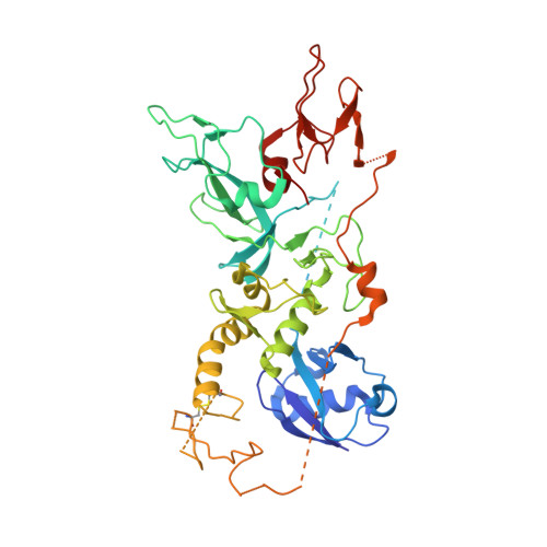
    4ZYN

  • PubMed Abstract: 

    Mutations in Parkin and PINK1 cause an inherited early-onset form of Parkinson's disease. The two proteins function together in a mitochondrial quality control pathway whereby PINK1 accumulates on damaged mitochondria and activates Parkin to induce mitophagy. How PINK1 kinase activity releases the auto-inhibited ubiquitin ligase activity of Parkin remains unclear. Here, we identify a binding switch between phospho-ubiquitin (pUb) and the ubiquitin-like domain (Ubl) of Parkin as a key element. By mutagenesis and SAXS, we show that pUb binds to RING1 of Parkin at a site formed by His302 and Arg305. pUb binding promotes disengagement of the Ubl from RING1 and subsequent Parkin phosphorylation. A crystal structure of Parkin Δ86-130 at 2.54 Å resolution allowed the design of mutations that specifically release the Ubl domain from RING1. These mutations mimic pUb binding and promote Parkin phosphorylation. Measurements of the E2 ubiquitin-conjugating enzyme UbcH7 binding to Parkin and Parkin E3 ligase activity suggest that Parkin phosphorylation regulates E3 ligase activity downstream of pUb binding.


  • Organizational Affiliation

    Groupe de recherché axé sur la structure des protéines and Department of Biochemistry, McGill University, Montréal, QC, Canada.


Macromolecules
Find similar proteins by:  (by identity cutoff)  |  3D Structure
Entity ID: 1
MoleculeChains Sequence LengthOrganismDetailsImage
E3 ubiquitin-protein ligase parkin
A, B
425Rattus norvegicusMutation(s): 0 
Gene Names: Park2Prkn
EC: 6.3.2
UniProt
Find proteins for Q9JK66 (Rattus norvegicus)
Explore Q9JK66 
Go to UniProtKB:  Q9JK66
Entity Groups  
Sequence Clusters30% Identity50% Identity70% Identity90% Identity95% Identity100% Identity
UniProt GroupQ9JK66
Sequence Annotations
Expand
  • Reference Sequence
Small Molecules
Ligands 2 Unique
IDChains Name / Formula / InChI Key2D Diagram3D Interactions
SO4
Query on SO4

Download Ideal Coordinates CCD File 
K [auth A]
L [auth A]
M [auth A]
N [auth A]
W [auth B]
K [auth A],
L [auth A],
M [auth A],
N [auth A],
W [auth B],
X [auth B]
SULFATE ION
O4 S
QAOWNCQODCNURD-UHFFFAOYSA-L
ZN
Query on ZN

Download Ideal Coordinates CCD File 
C [auth A]
D [auth A]
E [auth A]
F [auth A]
G [auth A]
C [auth A],
D [auth A],
E [auth A],
F [auth A],
G [auth A],
H [auth A],
I [auth A],
J [auth A],
O [auth B],
P [auth B],
Q [auth B],
R [auth B],
S [auth B],
T [auth B],
U [auth B],
V [auth B]
ZINC ION
Zn
PTFCDOFLOPIGGS-UHFFFAOYSA-N
Experimental Data & Validation

Experimental Data

  • Method: X-RAY DIFFRACTION
  • Resolution: 2.54 Å
  • R-Value Free: 0.235 
  • R-Value Work: 0.193 
  • R-Value Observed: 0.195 
  • Space Group: H 3
Unit Cell:
Length ( Å )Angle ( ˚ )
a = 114.105α = 90
b = 114.105β = 90
c = 186.399γ = 120
Software Package:
Software NamePurpose
REFMACrefinement
StructureStudiodata collection
PDB_EXTRACTdata extraction
XDSdata scaling
MOLREPphasing
XDSdata reduction

Structure Validation

View Full Validation Report


Entry History & Funding Information

Deposition Data


Funding OrganizationLocationGrant Number
Canadian Institutes of Health Research (CIHR)CanadaMOP-125924

Revision History  (Full details and data files)

  • Version 1.0: 2015-08-19
    Type: Initial release
  • Version 1.1: 2015-10-28
    Changes: Data collection, Database references
  • Version 1.2: 2017-09-20
    Changes: Author supporting evidence, Derived calculations
  • Version 1.3: 2020-01-08
    Changes: Author supporting evidence
  • Version 1.4: 2023-09-27
    Changes: Data collection, Database references, Refinement description