4ZYL

Crystal structure of response regulator RPA3017 in red light signaling of R. palustris


Experimental Data Snapshot

  • Method: X-RAY DIFFRACTION
  • Resolution: 1.80 Å
  • R-Value Free: 0.228 
  • R-Value Work: 0.189 
  • R-Value Observed: 0.190 

wwPDB Validation   3D Report Full Report


This is version 1.4 of the entry. See complete history


Literature

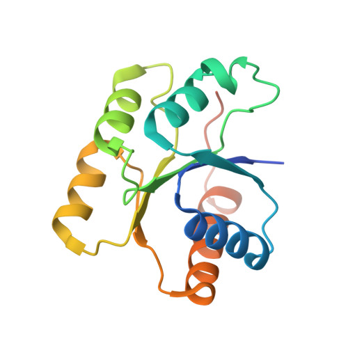
Structure of the response regulator RPA3017 involved in red-light signaling in Rhodopseudomonas palustris.

Yang, X.Zeng, X.Moffat, K.Yang, X.

(2015) Acta Crystallogr F Struct Biol Commun 71: 1215-1222

  • DOI: https://doi.org/10.1107/S2053230X15014661
  • Primary Citation of Related Structures:  
    4ZYL

  • PubMed Abstract: 

    Two-component signal transduction is the major signaling mechanism that enables bacteria to survive and thrive in complex environmental conditions. The photosynthetic bacterium R. palustris employs two tandem bacteriophytochromes, RpBphP2 and RpBphP3, to perceive red-light signals that regulate the synthesis of light-harvesting complexes under low-light conditions. Both RpBphP2 and RpBphP3 are photosensory histidine kinases coupled to the same response regulator RPA3017. Together, they constitute a two-component system that converts a red-light signal into a biological signal. In this work, the crystal structure of RPA3017 in the unphosphorylated form at 1.9 Å resolution is presented. This structure reveals a tightly associated dimer arrangement that is conserved among phytochrome-related response regulators. The conserved active-site architecture provides structural insight into the phosphotransfer reaction between RpBphP2/RpBphP3 and RPA3017. Based on structural comparisons and homology modeling, how specific recognition between RpBphP2/RpBphP3 and RPA3017 is achieved at the molecular level is further explored.


  • Organizational Affiliation

    National Key Laboratory of Crop Genetic Improvement, Huazhong Agricultural University, Wuhan 430070, People's Republic of China.


Macromolecules
Find similar proteins by:  (by identity cutoff)  |  3D Structure
Entity ID: 1
MoleculeChains Sequence LengthOrganismDetailsImage
RphyB protein
A, B
163Rhodopseudomonas palustris CGA009Mutation(s): 0 
Gene Names: rphyBRPA3017
UniProt
Find proteins for Q6N5G1 (Rhodopseudomonas palustris (strain ATCC BAA-98 / CGA009))
Explore Q6N5G1 
Go to UniProtKB:  Q6N5G1
Entity Groups  
Sequence Clusters30% Identity50% Identity70% Identity90% Identity95% Identity100% Identity
UniProt GroupQ6N5G1
Sequence Annotations
Expand
  • Reference Sequence
Experimental Data & Validation

Experimental Data

  • Method: X-RAY DIFFRACTION
  • Resolution: 1.80 Å
  • R-Value Free: 0.228 
  • R-Value Work: 0.189 
  • R-Value Observed: 0.190 
  • Space Group: P 65
Unit Cell:
Length ( Å )Angle ( ˚ )
a = 60.98α = 90
b = 60.98β = 90
c = 200.45γ = 120
Software Package:
Software NamePurpose
PHENIXrefinement
HKL-2000data reduction
HKL-2000data scaling
PHENIXphasing

Structure Validation

View Full Validation Report


Entry History & Funding Information

Deposition Data


Funding OrganizationLocationGrant Number
National Institutes of Health/National Eye Institute (NIH/NEI)United StatesR01EY024363

Revision History  (Full details and data files)

  • Version 1.0: 2015-10-14
    Type: Initial release
  • Version 1.1: 2017-09-20
    Changes: Author supporting evidence, Derived calculations
  • Version 1.2: 2018-10-10
    Changes: Data collection, Database references, Structure summary
  • Version 1.3: 2019-12-11
    Changes: Author supporting evidence, Structure summary
  • Version 1.4: 2024-03-06
    Changes: Data collection, Database references