4ZUL

Structure ALDH7A1 complexed with alpha-aminoadipate


Experimental Data Snapshot

  • Method: X-RAY DIFFRACTION
  • Resolution: 1.76 Å
  • R-Value Free: 0.206 
  • R-Value Work: 0.168 
  • R-Value Observed: 0.170 

wwPDB Validation   3D Report Full Report


Ligand Structure Quality Assessment 


This is version 2.0 of the entry. See complete history


Literature

Structural Basis of Substrate Recognition by Aldehyde Dehydrogenase 7A1.

Luo, M.Tanner, J.J.

(2015) Biochemistry 54: 5513-5522

  • DOI: https://doi.org/10.1021/acs.biochem.5b00754
  • Primary Citation of Related Structures:  
    4ZUK, 4ZUL, 4ZVW, 4ZVX, 4ZVY

  • PubMed Abstract: 

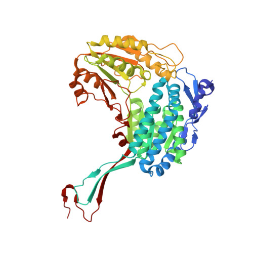
    Aldehyde dehydrogenase 7A1 (ALDH7A1) is part of lysine catabolism and catalyzes the NAD(+)-dependent oxidation of α-aminoadipate semialdehyde to α-aminoadipate. Herein, we describe a structural study of human ALDH7A1 focused on substrate recognition. Five crystal structures and small-angle X-ray scattering data are reported, including the first crystal structure of any ALDH7 family member complexed with α-aminoadipate. The product binds with the ε-carboxylate in the oxyanion hole, the aliphatic chain packed into an aromatic box, and the distal end of the product anchored by electrostatic interactions with five conserved residues. This binding mode resembles that of glutamate bound to the proline catabolic enzyme ALDH4A1. Analysis of ALDH7A1 and ALDH4A1 structures suggests key interactions that underlie substrate discrimination. Structures of apo ALDH7A1 reveal dramatic conformational differences from the product complex. Product binding is associated with a 16 Å movement of the C-terminus into the active site, which stabilizes the active conformation of the aldehyde substrate anchor loop. The fact that the C-terminus is part of the active site was hitherto unknown. Interestingly, the C-terminus and aldehyde anchor loop are disordered in a new tetragonal crystal form of the apoenzyme, implying that these parts of the enzyme are highly flexible. Our results suggest that the active site of ALDH7A1 is disassembled when the aldehyde site is vacant, and the C-terminus is a mobile element that forms quaternary structural interactions that aid aldehyde binding. These results are relevant to the c.1512delG genetic deletion associated with pyridoxine-dependent epilepsy, which alters the C-terminus of ALDH7A1.


  • Organizational Affiliation

    Department of Chemistry, University of Missouri-Columbia , Columbia, Missouri 65211, United States.


Macromolecules
Find similar proteins by:  (by identity cutoff)  |  3D Structure
Entity ID: 1
MoleculeChains Sequence LengthOrganismDetailsImage
Alpha-aminoadipic semialdehyde dehydrogenase
A, B, C, D, E
A, B, C, D, E, F, G, H
513Homo sapiensMutation(s): 0 
Gene Names: ALDH7A1ATQ1
EC: 1.2.1.31 (PDB Primary Data), 1.2.1.3 (PDB Primary Data), 1.2.1.8 (PDB Primary Data)
UniProt & NIH Common Fund Data Resources
Find proteins for P49419 (Homo sapiens)
Explore P49419 
Go to UniProtKB:  P49419
PHAROS:  P49419
GTEx:  ENSG00000164904 
Entity Groups  
Sequence Clusters30% Identity50% Identity70% Identity90% Identity95% Identity100% Identity
UniProt GroupP49419
Sequence Annotations
Expand
  • Reference Sequence
Small Molecules
Ligands 3 Unique
IDChains Name / Formula / InChI Key2D Diagram3D Interactions
PG4
Query on PG4

Download Ideal Coordinates CCD File 
Q [auth E],
W [auth H]
TETRAETHYLENE GLYCOL
C8 H18 O5
UWHCKJMYHZGTIT-UHFFFAOYSA-N
UN1
Query on UN1

Download Ideal Coordinates CCD File 
J [auth A]
L [auth B]
N [auth C]
P [auth D]
R [auth E]
J [auth A],
L [auth B],
N [auth C],
P [auth D],
R [auth E],
T [auth F],
V [auth G],
Y [auth H]
2-AMINOHEXANEDIOIC ACID
C6 H11 N O4
OYIFNHCXNCRBQI-BYPYZUCNSA-N
PGE
Query on PGE

Download Ideal Coordinates CCD File 
I [auth A]
K [auth B]
M [auth C]
O [auth D]
S [auth F]
I [auth A],
K [auth B],
M [auth C],
O [auth D],
S [auth F],
U [auth G],
X [auth H]
TRIETHYLENE GLYCOL
C6 H14 O4
ZIBGPFATKBEMQZ-UHFFFAOYSA-N
Experimental Data & Validation

Experimental Data

  • Method: X-RAY DIFFRACTION
  • Resolution: 1.76 Å
  • R-Value Free: 0.206 
  • R-Value Work: 0.168 
  • R-Value Observed: 0.170 
  • Space Group: C 1 2 1
Unit Cell:
Length ( Å )Angle ( ˚ )
a = 154.43α = 90
b = 162.534β = 95.04
c = 158.678γ = 90
Software Package:
Software NamePurpose
Aimlessdata scaling
PHENIXrefinement
PDB_EXTRACTdata extraction
XDSdata reduction
PHASERphasing

Structure Validation

View Full Validation Report


Ligand Structure Quality Assessment 


Entry History 

Deposition Data

Revision History  (Full details and data files)

  • Version 1.0: 2015-08-26
    Type: Initial release
  • Version 1.1: 2015-09-16
    Changes: Database references
  • Version 1.2: 2023-09-27
    Changes: Data collection, Database references, Derived calculations, Refinement description
  • Version 2.0: 2023-11-15
    Changes: Atomic model, Data collection