


Crystal structure of NADP-dependent dehydrogenase from Rhodobactersphaeroides in complex with NADP and sulfate
Kowiel, M., Gasiorowska, O.A., Shabalin, I.G., Handing, K.B., Porebski, P.J., Bonanno, J., Almo, S.C., Minor, W.To be published.
Experimental Data Snapshot
Entity ID: 1 | |||||
|---|---|---|---|---|---|
| Molecule | Chains | Sequence Length | Organism | Details | Image |
| NADP-dependent dehydrogenase | 316 | Cereibacter sphaeroides 2.4.1 | Mutation(s): 0 Gene Names: RSP_3442 EC: 1.1.1.79 (PDB Primary Data), 1.1.1.81 (PDB Primary Data) | 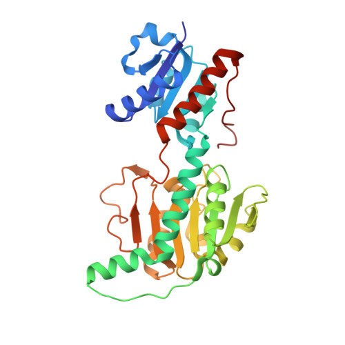 | |
UniProt | |||||
Find proteins for Q3IWN8 (Cereibacter sphaeroides (strain ATCC 17023 / DSM 158 / JCM 6121 / CCUG 31486 / LMG 2827 / NBRC 12203 / NCIMB 8253 / ATH 2.4.1.)) Explore Q3IWN8 Go to UniProtKB: Q3IWN8 | |||||
Entity Groups | |||||
| Sequence Clusters | 30% Identity50% Identity70% Identity90% Identity95% Identity100% Identity | ||||
| UniProt Group | Q3IWN8 | ||||
Sequence AnnotationsExpand | |||||
| |||||
| Ligands 5 Unique | |||||
|---|---|---|---|---|---|
| ID | Chains | Name / Formula / InChI Key | 2D Diagram | 3D Interactions | |
| NAP Query on NAP | C [auth A], F [auth B] | NADP NICOTINAMIDE-ADENINE-DINUCLEOTIDE PHOSPHATE C21 H28 N7 O17 P3 XJLXINKUBYWONI-NNYOXOHSSA-N |  | ||
| PGE Query on PGE | E [auth A] | TRIETHYLENE GLYCOL C6 H14 O4 ZIBGPFATKBEMQZ-UHFFFAOYSA-N |  | ||
| PEG Query on PEG | K [auth B] | DI(HYDROXYETHYL)ETHER C4 H10 O3 MTHSVFCYNBDYFN-UHFFFAOYSA-N |  | ||
| SO4 Query on SO4 | D [auth A], G [auth B], H [auth B], I [auth B] | SULFATE ION O4 S QAOWNCQODCNURD-UHFFFAOYSA-L |  | ||
| GOL Query on GOL | J [auth B] | GLYCEROL C3 H8 O3 PEDCQBHIVMGVHV-UHFFFAOYSA-N |  | ||
| Length ( Å ) | Angle ( ˚ ) |
|---|---|
| a = 71.489 | α = 90 |
| b = 71.489 | β = 90 |
| c = 244.364 | γ = 120 |
| Software Name | Purpose |
|---|---|
| PDB_EXTRACT | data extraction |
| Coot | model building |
| REFMAC | refinement |
| HKL-3000 | phasing |
| MOLREP | phasing |
| HKL-3000 | data scaling |
| HKL-3000 | data reduction |
| BLU-MAX | data collection |
| Funding Organization | Location | Grant Number |
|---|---|---|
| National Institutes of Health/National Institute of General Medical Sciences (NIH/NIGMS) | United States | -- |
RCSB PDB (citation) is hosted by
RCSB PDB is a member of the

