4ZAO

Genetically engineered Carbonic anhydrase IX


Experimental Data Snapshot

  • Method: X-RAY DIFFRACTION
  • Resolution: 1.80 Å
  • R-Value Free: 0.205 
  • R-Value Work: 0.175 
  • R-Value Observed: 0.177 

wwPDB Validation   3D Report Full Report


This is version 1.2 of the entry. See complete history


Literature

A sucrose-binding site provides a lead towards an isoform-specific inhibitor of the cancer-associated enzyme carbonic anhydrase IX.

Pinard, M.A.Aggarwal, M.Mahon, B.P.Tu, C.McKenna, R.

(2015) Acta Crystallogr F Struct Biol Commun 71: 1352-1358

  • DOI: https://doi.org/10.1107/S2053230X1501239X
  • Primary Citation of Related Structures:  
    4ZAO

  • PubMed Abstract: 

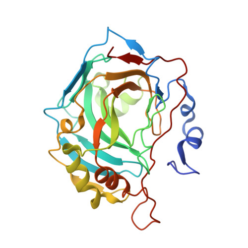
    Human carbonic anhydrase (CA; EC 4.2.1.1) isoform IX (CA IX) is an extracellular zinc metalloenzyme that catalyzes the reversible hydration of CO2 to HCO3(-), thereby playing a role in pH regulation. The majority of normal functioning cells exhibit low-level expression of CA IX. However, in cancer cells CA IX is upregulated as a consequence of a metabolic transition known as the Warburg effect. The upregulation of CA IX for cancer progression has drawn interest in it being a potential therapeutic target. CA IX is a transmembrane protein, and its purification, yield and crystallization have proven challenging to structure-based drug design, whereas the closely related cytosolic soluble isoform CA II can be expressed and crystallized with ease. Therefore, we have utilized structural alignments and site-directed mutagenesis to engineer a CA II that mimics the active site of CA IX. In this paper, the X-ray crystal structure of this CA IX mimic in complex with sucrose is presented and has been refined to a resolution of 1.5 Å, an Rcryst of 18.0% and an Rfree of 21.2%. The binding of sucrose at the entrance to the active site of the CA IX mimic, and not CA II, in a non-inhibitory mechanism provides a novel carbohydrate moiety binding site that could be further exploited to design isoform-specific inhibitors of CA IX.


  • Organizational Affiliation

    Department of Biochemistry and Molecular Biology, College of Medicine, University of Florida, Gainesville, FL 32610, USA.


Macromolecules
Find similar proteins by:  (by identity cutoff)  |  3D Structure
Entity ID: 1
MoleculeChains Sequence LengthOrganismDetailsImage
Carbonic anhydrase 2260Homo sapiensMutation(s): 7 
Gene Names: CA2
EC: 4.2.1.1
UniProt & NIH Common Fund Data Resources
Find proteins for P00918 (Homo sapiens)
Explore P00918 
Go to UniProtKB:  P00918
PHAROS:  P00918
GTEx:  ENSG00000104267 
Entity Groups  
Sequence Clusters30% Identity50% Identity70% Identity90% Identity95% Identity100% Identity
UniProt GroupP00918
Sequence Annotations
Expand
  • Reference Sequence
Small Molecules
Ligands 1 Unique
IDChains Name / Formula / InChI Key2D Diagram3D Interactions
ZN
Query on ZN

Download Ideal Coordinates CCD File 
B [auth A]ZINC ION
Zn
PTFCDOFLOPIGGS-UHFFFAOYSA-N
Experimental Data & Validation

Experimental Data

  • Method: X-RAY DIFFRACTION
  • Resolution: 1.80 Å
  • R-Value Free: 0.205 
  • R-Value Work: 0.175 
  • R-Value Observed: 0.177 
  • Space Group: P 1 21 1
Unit Cell:
Length ( Å )Angle ( ˚ )
a = 41.787α = 90
b = 41.231β = 103.83
c = 72.044γ = 90
Software Package:
Software NamePurpose
PHENIXrefinement
HKL-2000data reduction
HKL-2000data scaling
PHENIXphasing

Structure Validation

View Full Validation Report


Entry History 

Deposition Data

Revision History  (Full details and data files)

  • Version 1.0: 2015-10-14
    Type: Initial release
  • Version 1.1: 2018-04-18
    Changes: Advisory, Data collection, Database references, Derived calculations
  • Version 1.2: 2024-03-06
    Changes: Advisory, Data collection, Database references