4Z4P

Structure of the MLL4 SET Domain


Experimental Data Snapshot

  • Method: X-RAY DIFFRACTION
  • Resolution: 2.20 Å
  • R-Value Free: 0.242 
  • R-Value Work: 0.193 
  • R-Value Observed: 0.196 

wwPDB Validation   3D Report Full Report


Ligand Structure Quality Assessment 


This is version 1.2 of the entry. See complete history


Literature

Evolving Catalytic Properties of the MLL Family SET Domain.

Zhang, Y.Mittal, A.Reid, J.Reich, S.Gamblin, S.J.Wilson, J.R.

(2015) Structure 23: 1921-1933

  • DOI: https://doi.org/10.1016/j.str.2015.07.018
  • Primary Citation of Related Structures:  
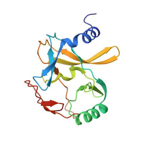
    4Z4P

  • PubMed Abstract: 

    Methylation of histone H3 lysine-4 is a hallmark of chromatin associated with active gene expression. The activity of H3K4-specific modification enzymes, in higher eukaryotes the MLL (or KMT2) family, is tightly regulated. The MLL family has six members, each with a specialized function. All contain a catalytic SET domain that associates with a core multiprotein complex for activation. These SET domains segregate into three classes that correlate with the arrangement of targeting domains that populate the rest of the protein. Here we show that, unlike MLL1, the MLL4 SET domain retains significant activity without the core complex. We also present the crystal structure of an inactive MLL4-tagged SET domain construct and describe conformational changes that account for MLL4 intrinsic activity. Finally, our structure explains how the MLL SET domains are able to add multiple methyl groups to the target lysine, despite having the sequence characteristics of a classical monomethylase.


  • Organizational Affiliation

    The Francis Crick Institute, Mill Hill Laboratory, London NW7 1AA, UK.


Macromolecules
Find similar proteins by:  (by identity cutoff)  |  3D Structure
Entity ID: 1
MoleculeChains Sequence LengthOrganismDetailsImage
Histone-lysine N-methyltransferase 2D166Homo sapiensMutation(s): 1 
Gene Names: KMT2DALRMLL2MLL4
EC: 2.1.1.43
UniProt & NIH Common Fund Data Resources
Find proteins for O14686 (Homo sapiens)
Explore O14686 
Go to UniProtKB:  O14686
PHAROS:  O14686
GTEx:  ENSG00000167548 
Entity Groups  
Sequence Clusters30% Identity50% Identity70% Identity90% Identity95% Identity100% Identity
UniProt GroupO14686
Sequence Annotations
Expand
  • Reference Sequence
Small Molecules
Ligands 2 Unique
IDChains Name / Formula / InChI Key2D Diagram3D Interactions
SAH
Query on SAH

Download Ideal Coordinates CCD File 
C [auth A]S-ADENOSYL-L-HOMOCYSTEINE
C14 H20 N6 O5 S
ZJUKTBDSGOFHSH-WFMPWKQPSA-N
ZN
Query on ZN

Download Ideal Coordinates CCD File 
B [auth A]ZINC ION
Zn
PTFCDOFLOPIGGS-UHFFFAOYSA-N
Experimental Data & Validation

Experimental Data

  • Method: X-RAY DIFFRACTION
  • Resolution: 2.20 Å
  • R-Value Free: 0.242 
  • R-Value Work: 0.193 
  • R-Value Observed: 0.196 
  • Space Group: P 1 21 1
Unit Cell:
Length ( Å )Angle ( ˚ )
a = 38.299α = 90
b = 40.692β = 109.78
c = 50.965γ = 90
Software Package:
Software NamePurpose
REFMACrefinement
XDSdata reduction
SCALAdata scaling
PHASERphasing

Structure Validation

View Full Validation Report


Ligand Structure Quality Assessment 


Entry History 

Deposition Data

Revision History  (Full details and data files)

  • Version 1.0: 2015-09-09
    Type: Initial release
  • Version 1.1: 2015-10-14
    Changes: Database references
  • Version 1.2: 2024-01-10
    Changes: Data collection, Database references, Refinement description