4Z31

Crystal structure of the RC3H2 ROQ domain in complex with stem-loop and double-stranded forms of RNA


Experimental Data Snapshot

  • Method: X-RAY DIFFRACTION
  • Resolution: 2.50 Å
  • R-Value Free: 0.259 
  • R-Value Work: 0.245 
  • R-Value Observed: 0.245 

wwPDB Validation   3D Report Full Report


This is version 1.2 of the entry. See complete history


Literature

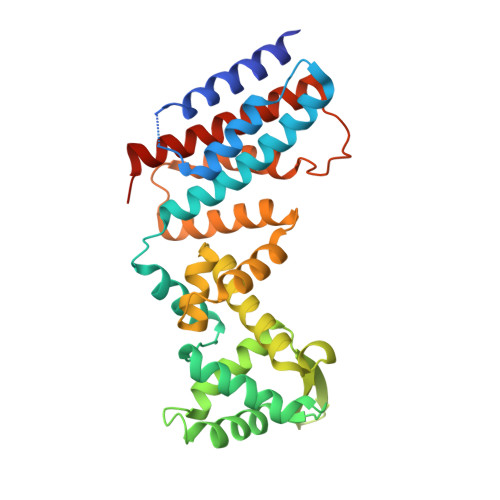
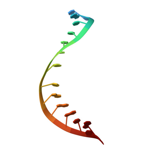
New Insights into the RNA-Binding and E3 Ubiquitin Ligase Activities of Roquins.

Zhang, Q.Fan, L.Hou, F.Dong, A.Wang, Y.X.Tong, Y.

(2015) Sci Rep 5: 15660-15660

  • DOI: https://doi.org/10.1038/srep15660
  • Primary Citation of Related Structures:  
    4Z30, 4Z31

  • PubMed Abstract: 

    Roquins are a family of highly conserved RNA-binding proteins that also contain a RING-type E3 ubiquitin ligase domain. They repress constitutive decay elements containing mRNAs and play a critical role in RNA homeostasis and immunological self-tolerance. Here we present the crystal structures of the RNA-binding region of Roquin paralog RC3H2 in both apo- and RNA-bound forms. The RNA-binding region has a bipartite architecture composed of ROQ and HEPN domains, and can bind to stem-loop and double-stranded RNAs simultaneously. The two domains undergo a large orientation change to accommodate RNA duplex binding. We profiled E2 ubiquitin-conjugating enzymes that pair with Roquins and found that RC3H1 and RC3H2 interact with two sets of overlapping but not identical E2 enzymes to drive the assembly of polyubiquitin chains of different linkages. Crystal structures, small-angle X-ray scattering, and E2 profiling revealed that while the two paralogs are highly homologous, RC3H2 and RC3H1 are different in their structures and functions. We also demonstrated that RNA duplex binding to RC3H2 cross-talks with its E3 ubiquitin ligase function using an in vitro auto-ubiquitination assay.


  • Organizational Affiliation

    Structural Genomics Consortium, University of Toronto, Toronto, ON M5G 1L7, Canada.


Macromolecules

Find similar proteins by:  (by identity cutoff)  |  3D Structure
Entity ID: 1
MoleculeChains Sequence LengthOrganismDetailsImage
Roquin-2
A, B
319Homo sapiensMutation(s): 0 
Gene Names: RC3H2MNABRNF164
UniProt & NIH Common Fund Data Resources
Find proteins for Q9HBD1 (Homo sapiens)
Explore Q9HBD1 
Go to UniProtKB:  Q9HBD1
PHAROS:  Q9HBD1
GTEx:  ENSG00000056586 
Entity Groups  
Sequence Clusters30% Identity50% Identity70% Identity90% Identity95% Identity100% Identity
UniProt GroupQ9HBD1
Sequence Annotations
Expand
  • Reference Sequence

Find similar nucleic acids by:  Sequence   |   3D Structure  

Entity ID: 2
MoleculeChains LengthOrganismImage
RNA (5'-R(*A)-D(P*UP*GP*UP*UP*CP*UP*GP*UP*GP*AP*AP*CP*AP*C)-3')
C, D, E, F
15Homo sapiens
Sequence Annotations
Expand
  • Reference Sequence
Experimental Data & Validation

Experimental Data

  • Method: X-RAY DIFFRACTION
  • Resolution: 2.50 Å
  • R-Value Free: 0.259 
  • R-Value Work: 0.245 
  • R-Value Observed: 0.245 
  • Space Group: P 1 21 1
Unit Cell:
Length ( Å )Angle ( ˚ )
a = 60.299α = 90
b = 174.734β = 114.12
c = 61.29γ = 90
Software Package:
Software NamePurpose
HKL-3000data scaling
REFMACrefinement
PDB_EXTRACTdata extraction
HKL-3000data reduction
PHASERphasing

Structure Validation

View Full Validation Report


Entry History 

Deposition Data

Revision History  (Full details and data files)

  • Version 1.0: 2015-10-14
    Type: Initial release
  • Version 1.1: 2016-06-29
    Changes: Database references
  • Version 1.2: 2023-09-27
    Changes: Data collection, Database references, Derived calculations, Refinement description