4YL6

Crystal structure of truncated cerebral cavernous malformation 2 C-terminal adaptor domain in complex with an internal helix of mitogen-activated protein kinase kinase kinase 3


Experimental Data Snapshot

  • Method: X-RAY DIFFRACTION
  • Resolution: 2.10 Å
  • R-Value Free: 0.237 
  • R-Value Work: 0.203 
  • R-Value Observed: 0.205 

wwPDB Validation   3D Report Full Report


This is version 1.2 of the entry. See complete history


Literature

Structural Insights into the Molecular Recognition between Cerebral Cavernous Malformation 2 and Mitogen-Activated Protein Kinase Kinase Kinase 3

Wang, X.Hou, Y.Deng, K.Zhang, Y.Wang, D.C.Ding, J.

(2015) Structure 23: 1087-1096

  • DOI: https://doi.org/10.1016/j.str.2015.04.003
  • Primary Citation of Related Structures:  
    4YKC, 4YKD, 4YL6

  • PubMed Abstract: 

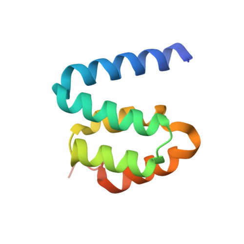
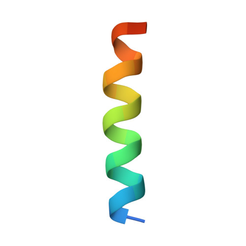
    Cerebral cavernous malformation 2 (CCM2) functions as an adaptor protein implicated in various biological processes. By interacting with the mitogen-activated protein kinase MEKK3, CCM2 either mediates the activation of MEKK3 signaling in response to osmotic stress or negatively regulates MEKK3 signaling, which is important for normal cardiovascular development. However, the molecular basis governing CCM2-MEKK3 interaction is largely unknown. Here we report the crystal structure of the CCM2 C-terminal part (CCM2ct) containing both the five-helix domain (CCM2cts) and the following C-terminal tail. The end of the C-terminal tail forms an isolated helix, which interacts intramolecularly with CCM2cts. By biochemical studies we identified the N-terminal amphiphilic helix of MEKK3 (MEKK3-nhelix) as the essential structural element for CCM2ct binding. We further determined the crystal structure of CCM2cts-MEKK3-nhelix complex, in which MEKK3-nhelix binds to the same site of CCM2cts for CCM2ct intramolecular interaction. These findings build a structural framework for understanding CCM2ct-MEKK3 molecular recognition.


  • Organizational Affiliation

    National Laboratory of Biomacromolecules, Institute of Biophysics, Chinese Academy of Sciences, Beijing 100101, People's Republic of China; Department of Clinical Oncology, Taihe Hospital, Hubei University of Medicine, Hubei 442000, People's Republic of China.


Macromolecules
Find similar proteins by:  (by identity cutoff)  |  3D Structure
Entity ID: 1
MoleculeChains Sequence LengthOrganismDetailsImage
Malcavernin96Homo sapiensMutation(s): 0 
Gene Names: CCM2
UniProt & NIH Common Fund Data Resources
Find proteins for Q9BSQ5 (Homo sapiens)
Explore Q9BSQ5 
Go to UniProtKB:  Q9BSQ5
PHAROS:  Q9BSQ5
GTEx:  ENSG00000136280 
Entity Groups  
Sequence Clusters30% Identity50% Identity70% Identity90% Identity95% Identity100% Identity
UniProt GroupQ9BSQ5
Sequence Annotations
Expand
  • Reference Sequence

Find similar proteins by:  Sequence   |   3D Structure  

Entity ID: 2
MoleculeChains Sequence LengthOrganismDetailsImage
Mitogen-activated protein kinase kinase kinase 322Homo sapiensMutation(s): 0 
UniProt & NIH Common Fund Data Resources
Find proteins for Q99759 (Homo sapiens)
Explore Q99759 
Go to UniProtKB:  Q99759
PHAROS:  Q99759
GTEx:  ENSG00000198909 
Entity Groups  
Sequence Clusters30% Identity50% Identity70% Identity90% Identity95% Identity100% Identity
UniProt GroupQ99759
Sequence Annotations
Expand
  • Reference Sequence
Experimental Data & Validation

Experimental Data

  • Method: X-RAY DIFFRACTION
  • Resolution: 2.10 Å
  • R-Value Free: 0.237 
  • R-Value Work: 0.203 
  • R-Value Observed: 0.205 
  • Space Group: P 43 21 2
Unit Cell:
Length ( Å )Angle ( ˚ )
a = 61.21α = 90
b = 61.21β = 90
c = 68.98γ = 90
Software Package:
Software NamePurpose
PHENIXrefinement
MOSFLMdata reduction
SCALAdata scaling
PHASERphasing

Structure Validation

View Full Validation Report


Entry History & Funding Information

Deposition Data


Funding OrganizationLocationGrant Number
Chinese Ministry of Science and Technology 973 programChina2011CB910304
Strategic Priority Research Program of the Chinese Academy of SciencesChinaXDB08020200

Revision History  (Full details and data files)

  • Version 1.0: 2015-06-03
    Type: Initial release
  • Version 1.1: 2015-06-17
    Changes: Database references
  • Version 1.2: 2023-11-08
    Changes: Data collection, Database references, Derived calculations, Refinement description, Source and taxonomy