4Y0F

Crystal Structure of Human TDP-43 RRM1 Domain in Complex with an Unmodified Single-stranded DNA


Experimental Data Snapshot

  • Method: X-RAY DIFFRACTION
  • Resolution: 2.65 Å
  • R-Value Free: 0.288 
  • R-Value Work: 0.240 
  • R-Value Observed: 0.245 

wwPDB Validation   3D Report Full Report


This is version 1.3 of the entry. See complete history


Literature

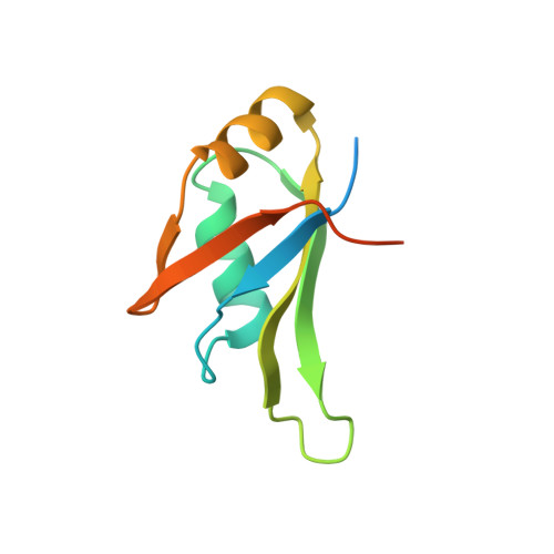
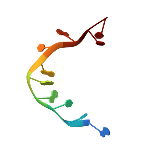
Structural analysis of disease-related TDP-43 D169G mutation: linking enhanced stability and caspase cleavage efficiency to protein accumulation

Chiang, C.H.Grauffel, C.Wu, L.S.Kuo, P.H.Doudeva, L.G.Lim, C.Shen, C.K.Yuan, H.S.

(2016) Sci Rep 6: 21581-21581

  • DOI: https://doi.org/10.1038/srep21581
  • Primary Citation of Related Structures:  
    4Y00, 4Y0F

  • PubMed Abstract: 

    The RNA-binding protein TDP-43 forms intracellular inclusions in amyotrophic lateral sclerosis (ALS). While TDP-43 mutations have been identified in ALS patients, how these mutations are linked to ALS remains unclear. Here we examined the biophysical properties of six ALS-linked TDP-43 mutants and found that one of the mutants, D169G, had higher thermal stability than wild-type TDP-43 and that it was cleaved by caspase 3 more efficiently, producing increased levels of the C-terminal 35 kD fragments (TDP-35) in vitro and in neuroblastoma cells. The crystal structure of the TDP-43 RRM1 domain containing the D169G mutation in complex with DNA along with molecular dynamics simulations reveal that the D169G mutation induces a local conformational change in a β turn and increases the hydrophobic interactions in the RRM1 core, thus enhancing the thermal stability of the RRM1 domain. Our results provide the first crystal structure of TDP-43 containing a disease-linked D169G mutation and a disease-related mechanism showing that D169G mutant is more susceptible to proteolytic cleavage by caspase 3 into the pathogenic C-terminal 35-kD fragments due to its increased stability in the RRM1 domain. Modulation of TDP-43 stability and caspase cleavage efficiency could present an avenue for prevention and treatment of TDP-43-linked neurodegeneration.


  • Organizational Affiliation

    Institute of Molecular Biology, Academia Sinica, Taipei, 11529, Taiwan.


Macromolecules

Find similar proteins by:  (by identity cutoff)  |  3D Structure
Entity ID: 1
MoleculeChains Sequence LengthOrganismDetailsImage
TAR DNA-binding protein 43
A, B
103Homo sapiensMutation(s): 0 
Gene Names: TARDBPTDP43
UniProt & NIH Common Fund Data Resources
Find proteins for Q13148 (Homo sapiens)
Explore Q13148 
Go to UniProtKB:  Q13148
PHAROS:  Q13148
GTEx:  ENSG00000120948 
Entity Groups  
Sequence Clusters30% Identity50% Identity70% Identity90% Identity95% Identity100% Identity
UniProt GroupQ13148
Sequence Annotations
Expand
  • Reference Sequence

Find similar nucleic acids by:  Sequence   |   3D Structure  

Entity ID: 2
MoleculeChains LengthOrganismImage
DNA (5'-D(*GP*TP*TP*GP*AP*GP*CP*GP*TP*T)-3')
C, D
10Homo sapiens
Sequence Annotations
Expand
  • Reference Sequence
Experimental Data & Validation

Experimental Data

  • Method: X-RAY DIFFRACTION
  • Resolution: 2.65 Å
  • R-Value Free: 0.288 
  • R-Value Work: 0.240 
  • R-Value Observed: 0.245 
  • Space Group: P 65 2 2
Unit Cell:
Length ( Å )Angle ( ˚ )
a = 100.027α = 90
b = 100.027β = 90
c = 98.144γ = 120
Software Package:
Software NamePurpose
PHENIXrefinement

Structure Validation

View Full Validation Report


Entry History & Funding Information

Deposition Data


Funding OrganizationLocationGrant Number
Academia SinicaTaiwan--
National Science CouncilTaiwan--

Revision History  (Full details and data files)

  • Version 1.0: 2016-02-10
    Type: Initial release
  • Version 1.1: 2016-03-09
    Changes: Database references
  • Version 1.2: 2017-10-04
    Changes: Derived calculations, Experimental preparation
  • Version 1.3: 2023-11-08
    Changes: Data collection, Database references, Refinement description