4XQ7

The crystal structure of the OAS-like domain (OLD) of human OASL


Experimental Data Snapshot

  • Method: X-RAY DIFFRACTION
  • Resolution: 1.60 Å
  • R-Value Free: 0.193 
  • R-Value Work: 0.167 
  • R-Value Observed: 0.168 

wwPDB Validation   3D Report Full Report


This is version 1.4 of the entry. See complete history


Literature

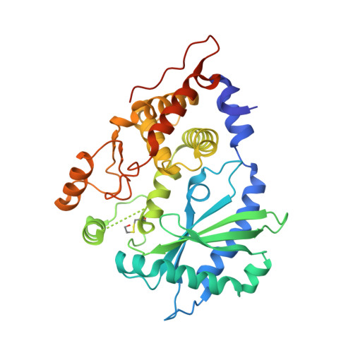
Structural and functional analysis reveals that human OASL binds dsRNA to enhance RIG-I signaling.

Ibsen, M.S.Gad, H.H.Andersen, L.L.Hornung, V.Julkunen, I.Sarkar, S.N.Hartmann, R.

(2015) Nucleic Acids Res 43: 5236-5248

  • DOI: https://doi.org/10.1093/nar/gkv389
  • Primary Citation of Related Structures:  
    4XQ7

  • PubMed Abstract: 

    The oligoadenylate synthetase (OAS) enzymes are cytoplasmic dsRNA sensors belonging to the antiviral innate immune system. Upon binding to viral dsRNA, the OAS enzymes synthesize 2'-5' linked oligoadenylates (2-5As) that initiate an RNA decay pathway to impair viral replication. The human OAS-like (OASL) protein, however, does not harbor the catalytic activity required for synthesizing 2-5As and differs from the other human OAS family members by having two C-terminal ubiquitin-like domains. In spite of its lack of enzymatic activity, human OASL possesses antiviral activity. It was recently demonstrated that the ubiquitin-like domains of OASL could substitute for K63-linked poly-ubiquitin and interact with the CARDs of RIG-I and thereby enhance RIG-I signaling. However, the role of the OAS-like domain of OASL remains unclear. Here we present the crystal structure of the OAS-like domain, which shows a striking similarity with activated OAS1. Furthermore, the structure of the OAS-like domain shows that OASL has a dsRNA binding groove. We demonstrate that the OAS-like domain can bind dsRNA and that mutating key residues in the dsRNA binding site is detrimental to the RIG-I signaling enhancement. Hence, binding to dsRNA is an important feature of OASL that is required for enhancing RIG-I signaling.


  • Organizational Affiliation

    Centre for Structural Biology, Department of Molecular Biology and Genetics, Aarhus University, 8000 Aarhus C, Denmark.


Macromolecules
Find similar proteins by:  (by identity cutoff)  |  3D Structure
Entity ID: 1
MoleculeChains Sequence LengthOrganismDetailsImage
2'-5'-oligoadenylate synthase-like protein359Homo sapiensMutation(s): 0 
Gene Names: OASLTRIP14
UniProt & NIH Common Fund Data Resources
Find proteins for Q15646 (Homo sapiens)
Explore Q15646 
Go to UniProtKB:  Q15646
PHAROS:  Q15646
GTEx:  ENSG00000135114 
Entity Groups  
Sequence Clusters30% Identity50% Identity70% Identity90% Identity95% Identity100% Identity
UniProt GroupQ15646
Sequence Annotations
Expand
  • Reference Sequence
Small Molecules
Modified Residues  1 Unique
IDChains TypeFormula2D DiagramParent
CME
Query on CME
A
L-PEPTIDE LINKINGC5 H11 N O3 S2CYS
Experimental Data & Validation

Experimental Data

  • Method: X-RAY DIFFRACTION
  • Resolution: 1.60 Å
  • R-Value Free: 0.193 
  • R-Value Work: 0.167 
  • R-Value Observed: 0.168 
  • Space Group: P 1 21 1
Unit Cell:
Length ( Å )Angle ( ˚ )
a = 54.8α = 90
b = 57.62β = 99.44
c = 64.79γ = 90
Software Package:
Software NamePurpose
PHENIXrefinement
XDSdata reduction
Cootmodel building
PHASERphasing
XSCALEdata scaling

Structure Validation

View Full Validation Report


Entry History 

Deposition Data

Revision History  (Full details and data files)

  • Version 1.0: 2015-04-22
    Type: Initial release
  • Version 1.1: 2015-05-13
    Changes: Database references
  • Version 1.2: 2015-06-10
    Changes: Database references
  • Version 1.3: 2018-01-17
    Changes: Data collection
  • Version 1.4: 2024-01-10
    Changes: Data collection, Database references, Refinement description