


Crystal structure of 3-hydroxyacyl-CoA dehydrogenase in complex with NAD from Burkholderia thailandensis
Abendroth, J., Dranow, D.M., Lorimer, D.D., Edwards, T.E.To be published.
Experimental Data Snapshot
Entity ID: 1 | |||||
|---|---|---|---|---|---|
| Molecule | Chains | Sequence Length | Organism | Details | Image |
| 3-hydroxyacyl-CoA dehydrogenase | 260 | Burkholderia thailandensis E264 | Mutation(s): 0 Gene Names: BTH_I0738 | 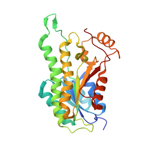 | |
UniProt | |||||
Find proteins for Q2T0K5 (Burkholderia thailandensis (strain ATCC 700388 / DSM 13276 / CCUG 48851 / CIP 106301 / E264)) Explore Q2T0K5 Go to UniProtKB: Q2T0K5 | |||||
Entity Groups | |||||
| Sequence Clusters | 30% Identity50% Identity70% Identity90% Identity95% Identity100% Identity | ||||
| UniProt Group | Q2T0K5 | ||||
Sequence AnnotationsExpand | |||||
| |||||
| Ligands 3 Unique | |||||
|---|---|---|---|---|---|
| ID | Chains | Name / Formula / InChI Key | 2D Diagram | 3D Interactions | |
| NAD Query on NAD | CA [auth G] GA [auth H] I [auth A] L [auth B] O [auth C] | NICOTINAMIDE-ADENINE-DINUCLEOTIDE C21 H27 N7 O14 P2 BAWFJGJZGIEFAR-NNYOXOHSSA-N |  | ||
| GOL Query on GOL | BA [auth G], FA [auth H], Q [auth C], R [auth D] | GLYCEROL C3 H8 O3 PEDCQBHIVMGVHV-UHFFFAOYSA-N |  | ||
| EDO Query on EDO | AA [auth F] DA [auth G] EA [auth G] HA [auth H] IA [auth H] | 1,2-ETHANEDIOL C2 H6 O2 LYCAIKOWRPUZTN-UHFFFAOYSA-N |  | ||
| Length ( Å ) | Angle ( ˚ ) |
|---|---|
| a = 71.22 | α = 103.26 |
| b = 77.1 | β = 93.98 |
| c = 109.35 | γ = 109.68 |
| Software Name | Purpose |
|---|---|
| XDS | data reduction |
| XSCALE | data scaling |
| PHASER | phasing |
| ARP | model building |
| Coot | model building |
| REFMAC | refinement |
| PDB_EXTRACT | data extraction |
RCSB PDB (citation) is hosted by
RCSB PDB is a member of the

