4XBH

Soluble rabbit neprilysin


Experimental Data Snapshot

  • Method: X-RAY DIFFRACTION
  • Resolution: 2.11 Å
  • R-Value Free: 0.211 
  • R-Value Work: 0.166 
  • R-Value Observed: 0.167 

wwPDB Validation   3D Report Full Report


Ligand Structure Quality Assessment 


This is version 2.1 of the entry. See complete history


Literature

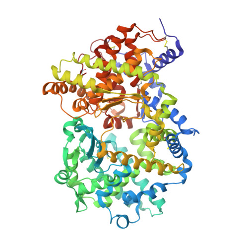
Structures of soluble rabbit neprilysin complexed with phosphoramidon or thiorphan.

Labiuk, S.L.Sygusch, J.Grochulski, P.

(2019) Acta Crystallogr F Struct Biol Commun 75: 405-411

  • DOI: https://doi.org/10.1107/S2053230X19006046
  • Primary Citation of Related Structures:  
    4XBH, 4ZR5, 5V48

  • PubMed Abstract: 

    Neutral endopeptidase (neprilysin; NEP) is a proteinase that cleaves a wide variety of peptides and has been implicated in Alzheimer's disease, cardiovascular conditions, arthritis and other inflammatory diseases. The structure of the soluble extracellular domain (residues 55-750) of rabbit neprilysin was solved both in its native form at 2.1 Å resolution, and bound to the inhibitors phosphoramidon and thiorphan at 2.8 and 3.0 Å resolution, respectively. Consistent with the extracellular domain of human neprilysin, the structure reveals a large central cavity which contains the active site and the location for inhibitor binding.


  • Organizational Affiliation

    Canadian Light Source, 44 Innovation Boulevard, Saskatoon, SK S7N 2V3, Canada.


Macromolecules
Find similar proteins by:  (by identity cutoff)  |  3D Structure
Entity ID: 1
MoleculeChains Sequence LengthOrganismDetailsImage
Neprilysin
A, B
696Oryctolagus cuniculusMutation(s): 0 
Gene Names: MME
EC: 3.4.24.11
UniProt
Find proteins for P08049 (Oryctolagus cuniculus)
Explore P08049 
Go to UniProtKB:  P08049
Entity Groups  
Sequence Clusters30% Identity50% Identity70% Identity90% Identity95% Identity100% Identity
UniProt GroupP08049
Sequence Annotations
Expand
  • Reference Sequence
Oligosaccharides

Help

Entity ID: 2
MoleculeChains Length2D Diagram Glycosylation3D Interactions
2-acetamido-2-deoxy-beta-D-glucopyranose-(1-4)-2-acetamido-2-deoxy-beta-D-glucopyranose
C, D, E, F
2N-Glycosylation
Glycosylation Resources
GlyTouCan:  G42666HT
GlyCosmos:  G42666HT
GlyGen:  G42666HT
Experimental Data & Validation

Experimental Data

  • Method: X-RAY DIFFRACTION
  • Resolution: 2.11 Å
  • R-Value Free: 0.211 
  • R-Value Work: 0.166 
  • R-Value Observed: 0.167 
  • Space Group: P 21 21 21
Unit Cell:
Length ( Å )Angle ( ˚ )
a = 69.361α = 90
b = 108.142β = 90
c = 212.784γ = 90
Software Package:
Software NamePurpose
HKL-2000data scaling
PHENIXrefinement
Cootmodel building
PDB_EXTRACTdata extraction

Structure Validation

View Full Validation Report


Ligand Structure Quality Assessment 


Entry History & Funding Information

Deposition Data


Funding OrganizationLocationGrant Number
Medical Research Council (MRC, Canada)Canada--

Revision History  (Full details and data files)

  • Version 1.0: 2016-05-25
    Type: Initial release
  • Version 1.1: 2017-09-20
    Changes: Author supporting evidence, Derived calculations, Refinement description
  • Version 1.2: 2019-06-26
    Changes: Data collection, Database references
  • Version 1.3: 2020-01-08
    Changes: Author supporting evidence
  • Version 2.0: 2020-07-29
    Type: Remediation
    Reason: Carbohydrate remediation
    Changes: Atomic model, Data collection, Derived calculations, Structure summary
  • Version 2.1: 2023-09-27
    Changes: Data collection, Database references, Refinement description, Structure summary