4XAU

Crystal structure of AtS13 from Actinomadura melliaura


Experimental Data Snapshot

  • Method: X-RAY DIFFRACTION
  • Resolution: 3.00 Å
  • R-Value Free: 0.220 
  • R-Value Work: 0.187 
  • R-Value Observed: 0.188 

wwPDB Validation   3D Report Full Report


Ligand Structure Quality Assessment 


This is version 1.6 of the entry. See complete history


Literature

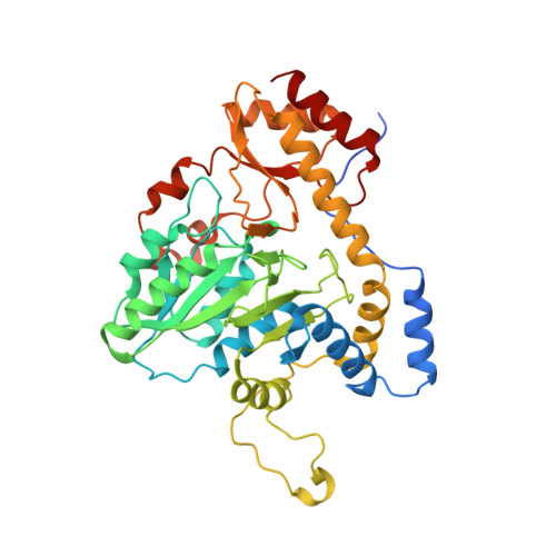
Structural characterization of AtmS13, a putative sugar aminotransferase involved in indolocarbazole AT2433 aminopentose biosynthesis.

Singh, S.Kim, Y.Wang, F.Bigelow, L.Endres, M.Kharel, M.K.Babnigg, G.Bingman, C.A.Joachimiak, A.Thorson, J.S.Phillips, G.N.

(2015) Proteins 83: 1547-1554

  • DOI: https://doi.org/10.1002/prot.24844
  • Primary Citation of Related Structures:  
    4XAU, 4ZWV

  • PubMed Abstract: 

    AT2433 from Actinomadura melliaura is an indolocarbazole antitumor antibiotic structurally distinguished by its unique aminodideoxypentose-containing disaccharide moiety. The corresponding sugar nucleotide-based biosynthetic pathway for this unusual sugar derives from comparative genomics where AtmS13 has been suggested as the contributing sugar aminotransferase (SAT). Determination of the AtmS13 X-ray structure at 1.50-Å resolution reveals it as a member of the aspartate aminotransferase fold type I (AAT-I). Structural comparisons of AtmS13 with homologous SATs that act upon similar substrates implicate potential active site residues that contribute to distinctions in sugar C5 (hexose vs. pentose) and/or sugar C2 (deoxy vs. hydroxyl) substrate specificity.


  • Organizational Affiliation

    Center for Pharmaceutical Research and Innovation, Pharmaceutical Sciences Division, University of Kentucky College of Pharmacy, Lexington, Kentucky, 40536-0596.


Macromolecules
Find similar proteins by:  (by identity cutoff)  |  3D Structure
Entity ID: 1
MoleculeChains Sequence LengthOrganismDetailsImage
Putative aminotransferase
A, B, C, D, E
A, B, C, D, E, F, G
389Actinomadura melliauraMutation(s): 0 
Gene Names: atS13
UniProt
Find proteins for Q0H2X1 (Actinomadura melliaura)
Explore Q0H2X1 
Go to UniProtKB:  Q0H2X1
Entity Groups  
Sequence Clusters30% Identity50% Identity70% Identity90% Identity95% Identity100% Identity
UniProt GroupQ0H2X1
Sequence Annotations
Expand
  • Reference Sequence
Small Molecules
Ligands 1 Unique
IDChains Name / Formula / InChI Key2D Diagram3D Interactions
PLP
Query on PLP

Download Ideal Coordinates CCD File 
H [auth C]PYRIDOXAL-5'-PHOSPHATE
C8 H10 N O6 P
NGVDGCNFYWLIFO-UHFFFAOYSA-N
Experimental Data & Validation

Experimental Data

  • Method: X-RAY DIFFRACTION
  • Resolution: 3.00 Å
  • R-Value Free: 0.220 
  • R-Value Work: 0.187 
  • R-Value Observed: 0.188 
  • Space Group: H 3 2
  • Diffraction Data: https://doi.org/10.18430/m34xau
Unit Cell:
Length ( Å )Angle ( ˚ )
a = 233.149α = 90
b = 233.149β = 90
c = 460.859γ = 120
Software Package:
Software NamePurpose
HKL-2000data reduction
PHENIXrefinement
PDB_EXTRACTdata extraction
HKL-2000data scaling
PHENIXphasing

Structure Validation

View Full Validation Report


Ligand Structure Quality Assessment 


Entry History & Funding Information

Deposition Data


Funding OrganizationLocationGrant Number
National Institutes of Health/National Institute of General Medical Sciences (NIH/NIGMS)United StatesU01GM098248

Revision History  (Full details and data files)

  • Version 1.0: 2014-12-24
    Type: Initial release
  • Version 1.1: 2015-06-24
    Changes: Database references
  • Version 1.2: 2015-07-29
    Changes: Database references
  • Version 1.3: 2015-08-26
    Changes: Data collection
  • Version 1.4: 2017-09-06
    Changes: Author supporting evidence, Derived calculations
  • Version 1.5: 2019-12-25
    Changes: Author supporting evidence
  • Version 1.6: 2023-09-27
    Changes: Data collection, Database references, Refinement description