4WUJ

Structural Biochemistry of a Fungal LOV Domain Photoreceptor Reveals an Evolutionarily Conserved Pathway Integrating Blue-Light and Oxidative Stress


Experimental Data Snapshot

  • Method: X-RAY DIFFRACTION
  • Resolution: 2.23 Å
  • R-Value Free: 0.260 
  • R-Value Work: 0.206 
  • R-Value Observed: 0.209 

wwPDB Validation   3D Report Full Report


Ligand Structure Quality Assessment 


This is version 1.3 of the entry. See complete history


Literature

Structural Biochemistry of a Fungal LOV Domain Photoreceptor Reveals an Evolutionarily Conserved Pathway Integrating Light and Oxidative Stress.

Lokhandwala, J.Hopkins, H.C.Rodriguez-Iglesias, A.Dattenbock, C.Schmoll, M.Zoltowski, B.D.

(2015) Structure 23: 116-125

  • DOI: https://doi.org/10.1016/j.str.2014.10.020
  • Primary Citation of Related Structures:  
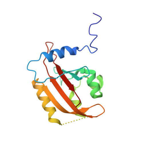
    4WUJ

  • PubMed Abstract: 

    Fungal LOV proteins facilitate photoadaptation via blue light regulation of dimer formation. Despite considerable homology of these proteins in closely related fungi, deviations in signaling exist. Here we report the crystal structure of ENVOY (ENV1), a homolog of N. crassa VVD in the fungus T. reesei, a model organism for plant cell wall degradation. Structural studies contradict a model of reversible competitive dimerization. Rather, evolutionary pressures have facilitated a two-residue shift in the position of a key Cys residue (Cys96) that enables the integration of environmental stress and light responses. A Cys96Thr variant abolishes adaptive responses to light and oxidative stress in a carbon source-dependent manner in vivo. Phylogenetic analysis verifies an evolutionary relevance of the Cys residue shift in different orders within Sordariomycetes. In this manner, we identified a widespread oxidative stress signaling mechanism that couples metabolic sensing and blue light responses not previously identified in LOV proteins.


  • Organizational Affiliation

    Department of Chemistry, Southern Methodist University, Dallas, TX 75275, USA.


Macromolecules
Find similar proteins by:  (by identity cutoff)  |  3D Structure
Entity ID: 1
MoleculeChains Sequence LengthOrganismDetailsImage
Glycoside hydrolase family 15, cellulose signaling associated protein envoy
A, B, C, D
147Trichoderma reesei QM6aMutation(s): 0 
Gene Names: env1TRIREDRAFT_81609
UniProt
Find proteins for G0RUC2 (Hypocrea jecorina (strain QM6a))
Explore G0RUC2 
Go to UniProtKB:  G0RUC2
Entity Groups  
Sequence Clusters30% Identity50% Identity70% Identity90% Identity95% Identity100% Identity
UniProt GroupG0RUC2
Sequence Annotations
Expand
  • Reference Sequence
Small Molecules
Ligands 2 Unique
IDChains Name / Formula / InChI Key2D Diagram3D Interactions
FMN
Query on FMN

Download Ideal Coordinates CCD File 
E [auth A],
G [auth B],
I [auth C],
L [auth D]
FLAVIN MONONUCLEOTIDE
C17 H21 N4 O9 P
FVTCRASFADXXNN-SCRDCRAPSA-N
SO4
Query on SO4

Download Ideal Coordinates CCD File 
F [auth A]
H [auth B]
J [auth C]
K [auth C]
M [auth D]
F [auth A],
H [auth B],
J [auth C],
K [auth C],
M [auth D],
N [auth D]
SULFATE ION
O4 S
QAOWNCQODCNURD-UHFFFAOYSA-L
Experimental Data & Validation

Experimental Data

  • Method: X-RAY DIFFRACTION
  • Resolution: 2.23 Å
  • R-Value Free: 0.260 
  • R-Value Work: 0.206 
  • R-Value Observed: 0.209 
  • Space Group: P 1 21 1
Unit Cell:
Length ( Å )Angle ( ˚ )
a = 44.342α = 90
b = 102.225β = 91.07
c = 71.55γ = 90
Software Package:
Software NamePurpose
HKL-3000phasing
Cootmodel building
PHASERphasing
PHENIXrefinement
PDB_EXTRACTdata extraction

Structure Validation

View Full Validation Report


Ligand Structure Quality Assessment 


Entry History & Funding Information

Deposition Data


Funding OrganizationLocationGrant Number
Herman Frasch FoundationUnited States739-HF12

Revision History  (Full details and data files)

  • Version 1.0: 2015-01-14
    Type: Initial release
  • Version 1.1: 2015-01-28
    Changes: Database references
  • Version 1.2: 2017-11-22
    Changes: Database references, Derived calculations, Other, Refinement description, Source and taxonomy
  • Version 1.3: 2023-09-27
    Changes: Data collection, Database references, Refinement description