4WTK

CRYSTAL STRUCTURE OF HCV NS5B GENOTYPE 2A JFH-1 ISOLATE WITH S15G E86Q E87Q C223H V321I MUTATIONS IN COMPLEX WITH RNA TEMPLATE 5'-AGCC, RNA PRIMER 5'-PGG, MN2+, AND CDP


Experimental Data Snapshot

  • Method: X-RAY DIFFRACTION
  • Resolution: 2.50 Å
  • R-Value Free: 0.224 
  • R-Value Work: 0.184 
  • R-Value Observed: 0.186 

wwPDB Validation   3D Report Full Report


Ligand Structure Quality Assessment 


This is version 1.3 of the entry. See complete history


Literature

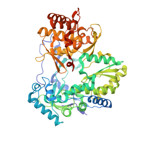
Structural basis for RNA replication by the hepatitis C virus polymerase.

Appleby, T.C.Perry, J.K.Murakami, E.Barauskas, O.Feng, J.Cho, A.Fox, D.Wetmore, D.R.McGrath, M.E.Ray, A.S.Sofia, M.J.Swaminathan, S.Edwards, T.E.

(2015) Science 347: 771-775

  • DOI: https://doi.org/10.1126/science.1259210
  • Primary Citation of Related Structures:  
    4WT9, 4WTA, 4WTC, 4WTD, 4WTE, 4WTF, 4WTG, 4WTI, 4WTJ, 4WTK, 4WTL, 4WTM

  • PubMed Abstract: 

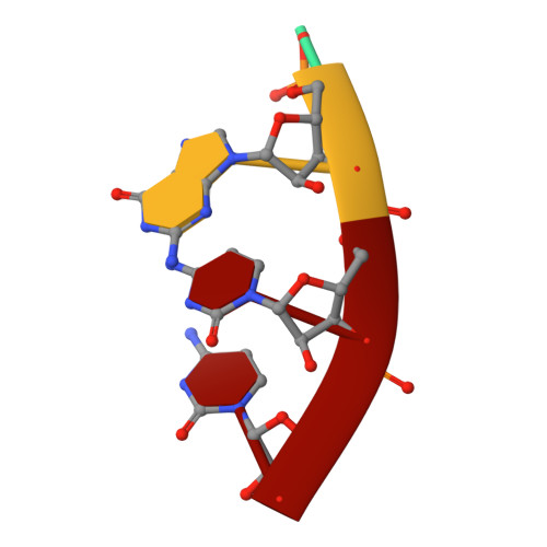
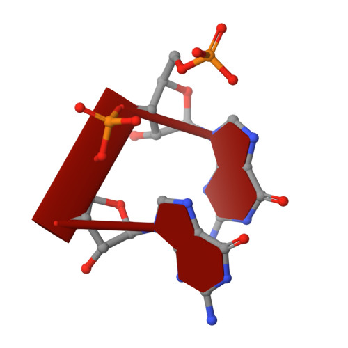
    Nucleotide analog inhibitors have shown clinical success in the treatment of hepatitis C virus (HCV) infection, despite an incomplete mechanistic understanding of NS5B, the viral RNA-dependent RNA polymerase. Here we study the details of HCV RNA replication by determining crystal structures of stalled polymerase ternary complexes with enzymes, RNA templates, RNA primers, incoming nucleotides, and catalytic metal ions during both primed initiation and elongation of RNA synthesis. Our analysis revealed that highly conserved active-site residues in NS5B position the primer for in-line attack on the incoming nucleotide. A β loop and a C-terminal membrane-anchoring linker occlude the active-site cavity in the apo state, retract in the primed initiation assembly to enforce replication of the HCV genome from the 3' terminus, and vacate the active-site cavity during elongation. We investigated the incorporation of nucleotide analog inhibitors, including the clinically active metabolite formed by sofosbuvir, to elucidate key molecular interactions in the active site.


  • Organizational Affiliation

    Gilead Sciences, 333 Lakeside Drive, Foster City, CA 94404, USA. todd.appleby@gilead.com tedwards@be4.com.


Macromolecules

Find similar proteins by:  (by identity cutoff)  |  3D Structure
Entity ID: 3
MoleculeChains Sequence LengthOrganismDetailsImage
RNA-directed RNA polymeraseC [auth A]580Hepatitis C virus JFH-1Mutation(s): 5 
EC: 2.7.7.48
UniProt
Find proteins for Q99IB8 (Hepatitis C virus genotype 2a (isolate JFH-1))
Explore Q99IB8 
Go to UniProtKB:  Q99IB8
Entity Groups  
Sequence Clusters30% Identity50% Identity70% Identity90% Identity95% Identity100% Identity
UniProt GroupQ99IB8
Sequence Annotations
Expand
  • Reference Sequence

Find similar nucleic acids by:  Sequence   |   3D Structure  

Entity ID: 1
MoleculeChains LengthOrganismImage
RNA TEMPLATE AGCCA [auth T]4synthetic construct
Sequence Annotations
Expand
  • Reference Sequence

Find similar nucleic acids by:  Sequence   |   3D Structure  

Entity ID: 2
MoleculeChains LengthOrganismImage
RNA PRIMER GGB [auth P]2synthetic construct
Sequence Annotations
Expand
  • Reference Sequence
Small Molecules
Ligands 6 Unique
IDChains Name / Formula / InChI Key2D Diagram3D Interactions
CDP
Query on CDP

Download Ideal Coordinates CCD File 
H [auth A]CYTIDINE-5'-DIPHOSPHATE
C9 H15 N3 O11 P2
ZWIADYZPOWUWEW-XVFCMESISA-N
B3P
Query on B3P

Download Ideal Coordinates CCD File 
J [auth A]2-[3-(2-HYDROXY-1,1-DIHYDROXYMETHYL-ETHYLAMINO)-PROPYLAMINO]-2-HYDROXYMETHYL-PROPANE-1,3-DIOL
C11 H26 N2 O6
HHKZCCWKTZRCCL-UHFFFAOYSA-N
PG6
Query on PG6

Download Ideal Coordinates CCD File 
I [auth A]1-(2-METHOXY-ETHOXY)-2-{2-[2-(2-METHOXY-ETHOXY]-ETHOXY}-ETHANE
C12 H26 O6
DMDPGPKXQDIQQG-UHFFFAOYSA-N
EDO
Query on EDO

Download Ideal Coordinates CCD File 
K [auth A]1,2-ETHANEDIOL
C2 H6 O2
LYCAIKOWRPUZTN-UHFFFAOYSA-N
MN
Query on MN

Download Ideal Coordinates CCD File 
D [auth A],
E [auth A],
F [auth A]
MANGANESE (II) ION
Mn
WAEMQWOKJMHJLA-UHFFFAOYSA-N
CL
Query on CL

Download Ideal Coordinates CCD File 
G [auth A]CHLORIDE ION
Cl
VEXZGXHMUGYJMC-UHFFFAOYSA-M
Experimental Data & Validation

Experimental Data

  • Method: X-RAY DIFFRACTION
  • Resolution: 2.50 Å
  • R-Value Free: 0.224 
  • R-Value Work: 0.184 
  • R-Value Observed: 0.186 
  • Space Group: P 65
Unit Cell:
Length ( Å )Angle ( ˚ )
a = 140.23α = 90
b = 140.23β = 90
c = 92.54γ = 120
Software Package:
Software NamePurpose
XSCALEdata scaling
REFMACrefinement
PDB_EXTRACTdata extraction
PHASERphasing

Structure Validation

View Full Validation Report


Ligand Structure Quality Assessment 


Entry History 

Deposition Data

Revision History  (Full details and data files)

  • Version 1.0: 2015-02-11
    Type: Initial release
  • Version 1.1: 2015-02-25
    Changes: Database references
  • Version 1.2: 2017-09-27
    Changes: Database references, Derived calculations, Other, Refinement description, Source and taxonomy
  • Version 1.3: 2023-09-27
    Changes: Data collection, Database references, Derived calculations, Refinement description