4W4Z

Structure of the EphA4 LBD in complex with peptide


Experimental Data Snapshot

  • Method: X-RAY DIFFRACTION
  • Resolution: 2.41 Å
  • R-Value Free: 0.241 
  • R-Value Work: 0.173 
  • R-Value Observed: 0.177 

wwPDB Validation   3D Report Full Report


This is version 1.5 of the entry. See complete history


Literature

Development and Structural Analysis of a Nanomolar Cyclic Peptide Antagonist for the EphA4 Receptor.

Lamberto, I.Lechtenberg, B.C.Olson, E.J.Mace, P.D.Dawson, P.E.Riedl, S.J.Pasquale, E.B.

(2014) ACS Chem Biol 9: 2787-2795

  • DOI: https://doi.org/10.1021/cb500677x
  • Primary Citation of Related Structures:  
    4W4Z, 4W50

  • PubMed Abstract: 

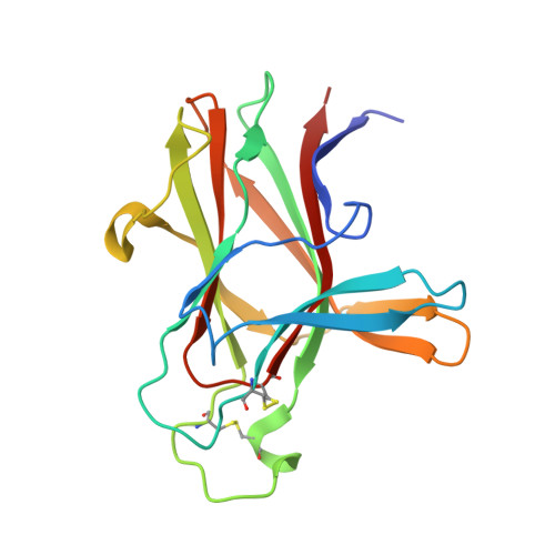
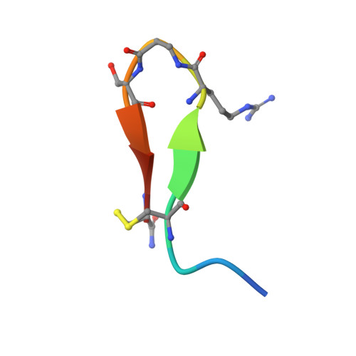
    The EphA4 receptor is highly expressed in the nervous system, and recent findings suggest that its signaling activity hinders neural repair and exacerbates certain neurodegenerative processes. EphA4 has also been implicated in cancer progression. Thus, EphA4 inhibitors represent potential therapeutic leads and useful research tools to elucidate the role of EphA4 in physiology and disease. Here, we report the structure of a cyclic peptide antagonist, APY, in complex with the EphA4 ligand-binding domain (LBD), which represents the first structure of a cyclic peptide bound to a receptor tyrosine kinase. The structure shows that the dodecameric APY efficiently occupies the ephrin ligand-binding pocket of EphA4 and promotes a "closed" conformation of the surrounding loops. Structure-guided relaxation of the strained APY β-turn and amidation of the C terminus to allow an additional intrapeptide hydrogen bond yielded APY-βAla8.am, an improved APY derivative that binds to EphA4 with nanomolar affinity. APY-βAla8.am potently inhibits ephrin-induced EphA4 activation in cells and EphA4-dependent neuronal growth cone collapse, while retaining high selectivity for EphA4. The two crystal structures of APY and APY-βAla8.am bound to EphA4, in conjunction with secondary phage display screens, highlighted peptide residues that are essential for EphA4 binding as well as residues that can be modified. Thus, the APY scaffold represents an exciting prototype, particularly since cyclic peptides have potentially favorable metabolic stability and are emerging as an important class of molecules for disruption of protein-protein interactions.


  • Organizational Affiliation

    Cancer Center, Sanford-Burnham Medical Research Institute , 10901 North Torrey Pines Road, La Jolla, California 92037, United States.


Macromolecules
Find similar proteins by:  (by identity cutoff)  |  3D Structure
Entity ID: 1
MoleculeChains Sequence LengthOrganismDetailsImage
Ephrin type-A receptor 4
A, B, C, D
179Homo sapiensMutation(s): 1 
Gene Names: EPHA4HEK8SEKTYRO1
EC: 2.7.10.1
UniProt & NIH Common Fund Data Resources
Find proteins for P54764 (Homo sapiens)
Explore P54764 
Go to UniProtKB:  P54764
PHAROS:  P54764
GTEx:  ENSG00000116106 
Entity Groups  
Sequence Clusters30% Identity50% Identity70% Identity90% Identity95% Identity100% Identity
UniProt GroupP54764
Sequence Annotations
Expand
  • Reference Sequence

Find similar proteins by:  Sequence   |   3D Structure  

Entity ID: 2
MoleculeChains Sequence LengthOrganismDetailsImage
APY-bAla8.am peptide
E, F, G, H
13synthetic constructMutation(s): 0 
Entity Groups  
Sequence Clusters30% Identity50% Identity70% Identity90% Identity95% Identity100% Identity
Sequence Annotations
Expand
  • Reference Sequence
Small Molecules
Ligands 2 Unique
IDChains Name / Formula / InChI Key2D Diagram3D Interactions
HEZ
Query on HEZ

Download Ideal Coordinates CCD File 
I [auth A]
J [auth A]
N [auth B]
O [auth B]
X [auth D]
I [auth A],
J [auth A],
N [auth B],
O [auth B],
X [auth D],
Y [auth D],
Z [auth D]
HEXANE-1,6-DIOL
C6 H14 O2
XXMIOPMDWAUFGU-UHFFFAOYSA-N
GOL
Query on GOL

Download Ideal Coordinates CCD File 
AA [auth D]
BA [auth D]
CA [auth H]
K [auth A]
L [auth A]
AA [auth D],
BA [auth D],
CA [auth H],
K [auth A],
L [auth A],
M [auth A],
P [auth B],
Q [auth B],
R [auth B],
S [auth B],
T [auth C],
U [auth C],
V [auth C],
W [auth C]
GLYCEROL
C3 H8 O3
PEDCQBHIVMGVHV-UHFFFAOYSA-N
Experimental Data & Validation

Experimental Data

  • Method: X-RAY DIFFRACTION
  • Resolution: 2.41 Å
  • R-Value Free: 0.241 
  • R-Value Work: 0.173 
  • R-Value Observed: 0.177 
  • Space Group: P 1 21 1
Unit Cell:
Length ( Å )Angle ( ˚ )
a = 37.22α = 90
b = 127.19β = 90
c = 84.599γ = 90
Software Package:
Software NamePurpose
CrystalCleardata collection
MOSFLMdata reduction
Aimlessdata scaling
REFMACrefinement
Cootmodel building
PDB_EXTRACTdata extraction

Structure Validation

View Full Validation Report


Entry History & Funding Information

Deposition Data


Funding OrganizationLocationGrant Number
National Institutes of Health/National Cancer Institute (NIH/NCI)United StatesP01CA138390

Revision History  (Full details and data files)

  • Version 1.0: 2014-10-08
    Type: Initial release
  • Version 1.1: 2015-01-07
    Changes: Database references
  • Version 1.2: 2017-09-06
    Changes: Advisory, Author supporting evidence, Derived calculations, Other, Source and taxonomy, Structure summary
  • Version 1.3: 2017-09-27
    Changes: Data collection, Refinement description
  • Version 1.4: 2019-12-04
    Changes: Author supporting evidence
  • Version 1.5: 2023-12-27
    Changes: Data collection, Database references, Refinement description