4V2X

High resolution structure of the full length tri-modular endo-beta-1, 4-glucanase B (Cel5B) from Bacillus halodurans


Experimental Data Snapshot

  • Method: X-RAY DIFFRACTION
  • Resolution: 1.64 Å
  • R-Value Free: 0.181 
  • R-Value Work: 0.154 
  • R-Value Observed: 0.156 

wwPDB Validation   3D Report Full Report


This is version 1.3 of the entry. See complete history


Literature

Family 46 Carbohydrate-Binding Modules Contribute to the Enzymatic Hydrolysis of Xyloglucan and Beta-1,3-1,4-Glucans Through Distinct Mechanisms.

Venditto, I.Najmudin, S.Luis, A.S.Ferreira, L.M.Sakka, K.Knox, J.P.Gilbert, H.J.Fontes, C.M.

(2015) J Biol Chem 290: 10572

  • DOI: https://doi.org/10.1074/jbc.M115.637827
  • Primary Citation of Related Structures:  
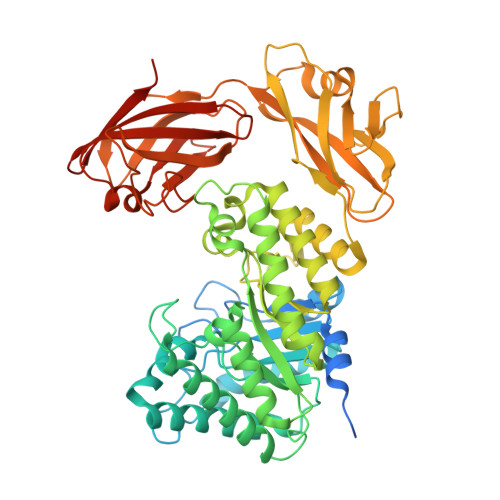
    4UZ8, 4UZN, 4V2X

  • PubMed Abstract: 

    Structural carbohydrates comprise an extraordinary source of energy that remains poorly utilized by the biofuel sector as enzymes have restricted access to their substrates within the intricacy of plant cell walls. Carbohydrate active enzymes (CAZYmes) that target recalcitrant polysaccharides are modular enzymes containing noncatalytic carbohydrate-binding modules (CBMs) that direct enzymes to their cognate substrate, thus potentiating catalysis. In general, CBMs are functionally and structurally autonomous from their associated catalytic domains from which they are separated through flexible linker sequences. Here, we show that a C-terminal CBM46 derived from BhCel5B, a Bacillus halodurans endoglucanase, does not interact with β-glucans independently but, uniquely, acts cooperatively with the catalytic domain of the enzyme in substrate recognition. The structure of BhCBM46 revealed a β-sandwich fold that abuts onto the region of the substrate binding cleft upstream of the active site. BhCBM46 as a discrete entity is unable to bind to β-glucans. Removal of BhCBM46 from BhCel5B, however, abrogates binding to β-1,3-1,4-glucans while substantially decreasing the affinity for decorated β-1,4-glucan homopolymers such as xyloglucan. The CBM46 was shown to contribute to xyloglucan hydrolysis only in the context of intact plant cell walls, but it potentiates enzymatic activity against purified β-1,3-1,4-glucans in solution or within the cell wall. This report reveals the mechanism by which a CBM can promote enzyme activity through direct interaction with the substrate or by targeting regions of the plant cell wall where the target glucan is abundant.


  • Organizational Affiliation

    From the Centro Interdisciplinar de Investigação em Sanidade Animal, Faculdade de Medicina Veterinária, ULisboa, Pólo Universitário do Alto da Ajuda, Avenida da Universidade Técnica, 1300-477 Lisboa, Portugal.


Macromolecules
Find similar proteins by:  (by identity cutoff)  |  3D Structure
Entity ID: 1
MoleculeChains Sequence LengthOrganismDetailsImage
ENDO-BETA-1,4-GLUCANASE (CELLULASE B)597Halalkalibacterium haloduransMutation(s): 0 
UniProt
Find proteins for Q9KF82 (Halalkalibacterium halodurans (strain ATCC BAA-125 / DSM 18197 / FERM 7344 / JCM 9153 / C-125))
Explore Q9KF82 
Go to UniProtKB:  Q9KF82
Entity Groups  
Sequence Clusters30% Identity50% Identity70% Identity90% Identity95% Identity100% Identity
UniProt GroupQ9KF82
Sequence Annotations
Expand
  • Reference Sequence
Small Molecules
Ligands 4 Unique
IDChains Name / Formula / InChI Key2D Diagram3D Interactions
CAC
Query on CAC

Download Ideal Coordinates CCD File 
K [auth A]CACODYLATE ION
C2 H6 As O2
OGGXGZAMXPVRFZ-UHFFFAOYSA-M
GOL
Query on GOL

Download Ideal Coordinates CCD File 
E [auth A]
F [auth A]
G [auth A]
H [auth A]
I [auth A]
E [auth A],
F [auth A],
G [auth A],
H [auth A],
I [auth A],
J [auth A]
GLYCEROL
C3 H8 O3
PEDCQBHIVMGVHV-UHFFFAOYSA-N
ACT
Query on ACT

Download Ideal Coordinates CCD File 
D [auth A]ACETATE ION
C2 H3 O2
QTBSBXVTEAMEQO-UHFFFAOYSA-M
CA
Query on CA

Download Ideal Coordinates CCD File 
B [auth A],
C [auth A]
CALCIUM ION
Ca
BHPQYMZQTOCNFJ-UHFFFAOYSA-N
Experimental Data & Validation

Experimental Data

  • Method: X-RAY DIFFRACTION
  • Resolution: 1.64 Å
  • R-Value Free: 0.181 
  • R-Value Work: 0.154 
  • R-Value Observed: 0.156 
  • Space Group: P 21 21 2
Unit Cell:
Length ( Å )Angle ( ˚ )
a = 74.22α = 90
b = 141.84β = 90
c = 50.82γ = 90
Software Package:
Software NamePurpose
REFMACrefinement
XDSdata reduction
xia2data scaling
BALBESphasing

Structure Validation

View Full Validation Report


Entry History 

Deposition Data

Revision History  (Full details and data files)

  • Version 1.0: 2015-02-25
    Type: Initial release
  • Version 1.1: 2015-03-11
    Changes: Database references
  • Version 1.2: 2015-05-13
    Changes: Database references
  • Version 1.3: 2024-01-10
    Changes: Data collection, Database references, Derived calculations, Other, Refinement description