Enantioselective Oxidation of Galactitol 1-Phosphate by Galactitol-1-Phosphate 5-Dehydrogenase from Escherichia Coli
Benavente, R., Esteban-Torres, M., Kohring, G.W., Cortes-Cabrera, A., Sanchez-Murcia, P.A., Gago, F., Acebron, I., De Las Rivas, B., Munoz, R., Mancheno, J.M.(2015) Acta Crystallogr D Biol Crystallogr 71: 1540
- PubMed: 26143925
- DOI: https://doi.org/10.1107/S1399004715009281
- Primary Citation of Related Structures:
4UEJ, 4UEK, 4UEO - PubMed Abstract:
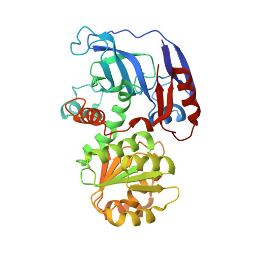
Galactitol-1-phosphate 5-dehydrogenase (GPDH) is a polyol dehydrogenase that belongs to the medium-chain dehydrogenase/reductase (MDR) superfamily. It catalyses the Zn(2+)- and NAD(+)-dependent stereoselective dehydrogenation of L-galactitol 1-phosphate to D-tagatose 6-phosphate. Here, three crystal structures of GPDH from Escherichia coli are reported: that of the open state of GPDH with Zn(2+) in the catalytic site and those of the closed state in complex with the polyols Tris and glycerol, respectively. The closed state of GPDH reveals no bound cofactor, which is at variance with the conformational transition of the prototypical mammalian liver alcohol dehydrogenase. The main intersubunit-contacting interface within the GPDH homodimer presents a large internal cavity that probably facilitates the relative movement between the subunits. The substrate analogue glycerol bound within the active site partially mimics the catalytically relevant backbone of galactitol 1-phosphate. The glycerol binding mode reveals, for the first time in the polyol dehydrogenases, a pentacoordinated zinc ion in complex with a polyol and also a strong hydrogen bond between the primary hydroxyl group and the conserved Glu144, an interaction originally proposed more than thirty years ago that supports a catalytic role for this acidic residue.
Organizational Affiliation:
Department of Crystallography and Structural Biology, Institute of Physical Chemistry Rocasolano, CSIC, Serrano 119, 28006 Madrid, Spain.