4U7D

Structure of human RECQ-like helicase in complex with an oligonucleotide


Experimental Data Snapshot

  • Method: X-RAY DIFFRACTION
  • Resolution: 3.40 Å
  • R-Value Free: 0.249 
  • R-Value Work: 0.205 
  • R-Value Observed: 0.214 

wwPDB Validation   3D Report Full Report


This is version 1.4 of the entry. See complete history


Literature

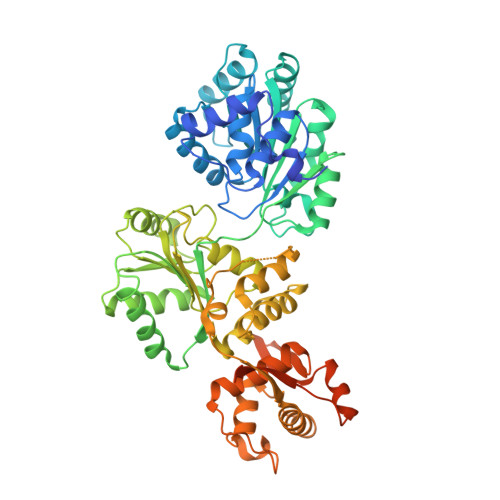
Human RECQ1 helicase-driven DNA unwinding, annealing, and branch migration: Insights from DNA complex structures.

Pike, A.C.Gomathinayagam, S.Swuec, P.Berti, M.Zhang, Y.Schnecke, C.Marino, F.von Delft, F.Renault, L.Costa, A.Gileadi, O.Vindigni, A.

(2015) Proc Natl Acad Sci U S A 112: 4286-4291

  • DOI: https://doi.org/10.1073/pnas.1417594112
  • Primary Citation of Related Structures:  
    2WWY, 4U7D

  • PubMed Abstract: 

    RecQ helicases are a widely conserved family of ATP-dependent motors with diverse roles in nearly every aspect of bacterial and eukaryotic genome maintenance. However, the physical mechanisms by which RecQ helicases recognize and process specific DNA replication and repair intermediates are largely unknown. Here, we solved crystal structures of the human RECQ1 helicase in complexes with tailed-duplex DNA and ssDNA. The structures map the interactions of the ssDNA tail and the branch point along the helicase and Zn-binding domains, which, together with reported structures of other helicases, define the catalytic stages of helicase action. We also identify a strand-separating pin, which (uniquely in RECQ1) is buttressed by the protein dimer interface. A duplex DNA-binding surface on the C-terminal domain is shown to play a role in DNA unwinding, strand annealing, and Holliday junction (HJ) branch migration. We have combined EM and analytical ultracentrifugation approaches to show that RECQ1 can form what appears to be a flat, homotetrameric complex and propose that RECQ1 tetramers are involved in HJ recognition. This tetrameric arrangement suggests a platform for coordinated activity at the advancing and receding duplexes of an HJ during branch migration.


  • Organizational Affiliation

    Structural Genomics Consortium, University of Oxford, Oxford OX3 7DQ, United Kingdom;


Macromolecules

Find similar proteins by:  (by identity cutoff)  |  3D Structure
Entity ID: 1
MoleculeChains Sequence LengthOrganismDetailsImage
ATP-dependent DNA helicase Q1
A, B, C, D
591Homo sapiensMutation(s): 0 
Gene Names: RECQLRECQ1RECQL1
EC: 3.6.4.12
UniProt & NIH Common Fund Data Resources
Find proteins for P46063 (Homo sapiens)
Explore P46063 
Go to UniProtKB:  P46063
PHAROS:  P46063
GTEx:  ENSG00000004700 
Entity Groups  
Sequence Clusters30% Identity50% Identity70% Identity90% Identity95% Identity100% Identity
UniProt GroupP46063
Sequence Annotations
Expand
  • Reference Sequence

Find similar nucleic acids by:  Sequence   |   3D Structure  

Entity ID: 2
MoleculeChains LengthOrganismImage
DNA oligonucleotideE [auth P],
F [auth Q],
G [auth R],
H [auth S]
20synthetic construct
Sequence Annotations
Expand
  • Reference Sequence
Experimental Data & Validation

Experimental Data

  • Method: X-RAY DIFFRACTION
  • Resolution: 3.40 Å
  • R-Value Free: 0.249 
  • R-Value Work: 0.205 
  • R-Value Observed: 0.214 
  • Space Group: P 1 21 1
Unit Cell:
Length ( Å )Angle ( ˚ )
a = 55.38α = 90
b = 138.22β = 90.02
c = 207.581γ = 90
Software Package:
Software NamePurpose
PHENIXrefinement
MOSFLMdata reduction
SCALAdata scaling
PHASERphasing

Structure Validation

View Full Validation Report


Entry History & Funding Information

Deposition Data


Funding OrganizationLocationGrant Number
Wellcome TrustUnited Kingdom092809/Z/10/Z

Revision History  (Full details and data files)

  • Version 1.0: 2015-01-21
    Type: Initial release
  • Version 1.1: 2015-04-01
    Changes: Database references
  • Version 1.2: 2015-04-15
    Changes: Database references
  • Version 1.3: 2018-01-24
    Changes: Structure summary
  • Version 1.4: 2023-12-20
    Changes: Data collection, Database references, Refinement description