4U46

Crystal structure of an avidin mutant


Experimental Data Snapshot

  • Method: X-RAY DIFFRACTION
  • Resolution: 1.95 Å
  • R-Value Free: 0.214 
  • R-Value Work: 0.182 
  • R-Value Observed: 0.184 

wwPDB Validation   3D Report Full Report


This is version 1.2 of the entry. See complete history


Literature

Artificial Avidin-Based Receptors for a Panel of Small Molecules.

Lehtonen, S.I.Tullila, A.Agrawal, N.Kukkurainen, S.Kahkonen, N.Koskinen, M.Nevanen, T.K.Johnson, M.S.Airenne, T.T.Kulomaa, M.S.Riihimaki, T.A.Hytonen, V.P.

(2016) ACS Chem Biol 11: 211-221

  • DOI: https://doi.org/10.1021/acschembio.5b00906
  • Primary Citation of Related Structures:  
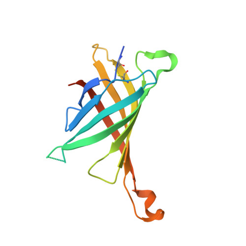
    4U46

  • PubMed Abstract: 

    Proteins with high specificity, affinity, and stability are needed for biomolecular recognition in a plethora of applications. Antibodies are powerful affinity tools, but they may also suffer from limitations such as low stability and high production costs. Avidin and streptavidin provide a promising scaffold for protein engineering, and due to their ultratight binding to D-biotin they are widely used in various biotechnological and biomedical applications. In this study, we demonstrate that the avidin scaffold is suitable for use as a novel receptor for several biologically active small molecules: Artificial, chicken avidin-based proteins, antidins, were generated using a directed evolution method for progesterone, hydrocortisone, testosterone, cholic acid, ketoprofen, and folic acid, all with micromolar to nanomolar affinity and significantly reduced biotin-binding affinity. We also describe the crystal structure of an antidin, sbAvd-2(I117Y), a steroid-binding avidin, which proves that the avidin scaffold can tolerate significant modifications without losing its characteristic tetrameric beta-barrel structure, helping us to further design avidin-based small molecule receptors.


  • Organizational Affiliation

    BioMediTech, University of Tampere , Biokatu 6, FI-33014 Tampere, Finland.


Macromolecules
Find similar proteins by:  (by identity cutoff)  |  3D Structure
Entity ID: 1
MoleculeChains Sequence LengthOrganismDetailsImage
Avidin
A, B
138Gallus gallusMutation(s): 0 
Gene Names: AVD
UniProt
Find proteins for P02701 (Gallus gallus)
Explore P02701 
Go to UniProtKB:  P02701
Entity Groups  
Sequence Clusters30% Identity50% Identity70% Identity90% Identity95% Identity100% Identity
UniProt GroupP02701
Sequence Annotations
Expand
  • Reference Sequence
Small Molecules
Ligands 1 Unique
IDChains Name / Formula / InChI Key2D Diagram3D Interactions
CL
Query on CL

Download Ideal Coordinates CCD File 
C [auth A]
D [auth A]
E [auth A]
F [auth A]
G [auth A]
C [auth A],
D [auth A],
E [auth A],
F [auth A],
G [auth A],
H [auth A],
I [auth B],
J [auth B],
K [auth B],
L [auth B],
M [auth B],
N [auth B]
CHLORIDE ION
Cl
VEXZGXHMUGYJMC-UHFFFAOYSA-M
Experimental Data & Validation

Experimental Data

  • Method: X-RAY DIFFRACTION
  • Resolution: 1.95 Å
  • R-Value Free: 0.214 
  • R-Value Work: 0.182 
  • R-Value Observed: 0.184 
  • Space Group: P 21 21 2
Unit Cell:
Length ( Å )Angle ( ˚ )
a = 74.48α = 90
b = 79.8β = 90
c = 43.07γ = 90
Software Package:
Software NamePurpose
XDSdata reduction
REFMACrefinement
XSCALEdata scaling
PHASERphasing

Structure Validation

View Full Validation Report


Entry History 

Deposition Data

Revision History  (Full details and data files)

  • Version 1.0: 2015-08-05
    Type: Initial release
  • Version 1.1: 2016-09-28
    Changes: Database references
  • Version 1.2: 2023-12-20
    Changes: Data collection, Database references, Refinement description