4U3C

Docking Site of Maltohexaose in the Mtb GlgE


Experimental Data Snapshot

  • Method: X-RAY DIFFRACTION
  • Resolution: 3.98 Å
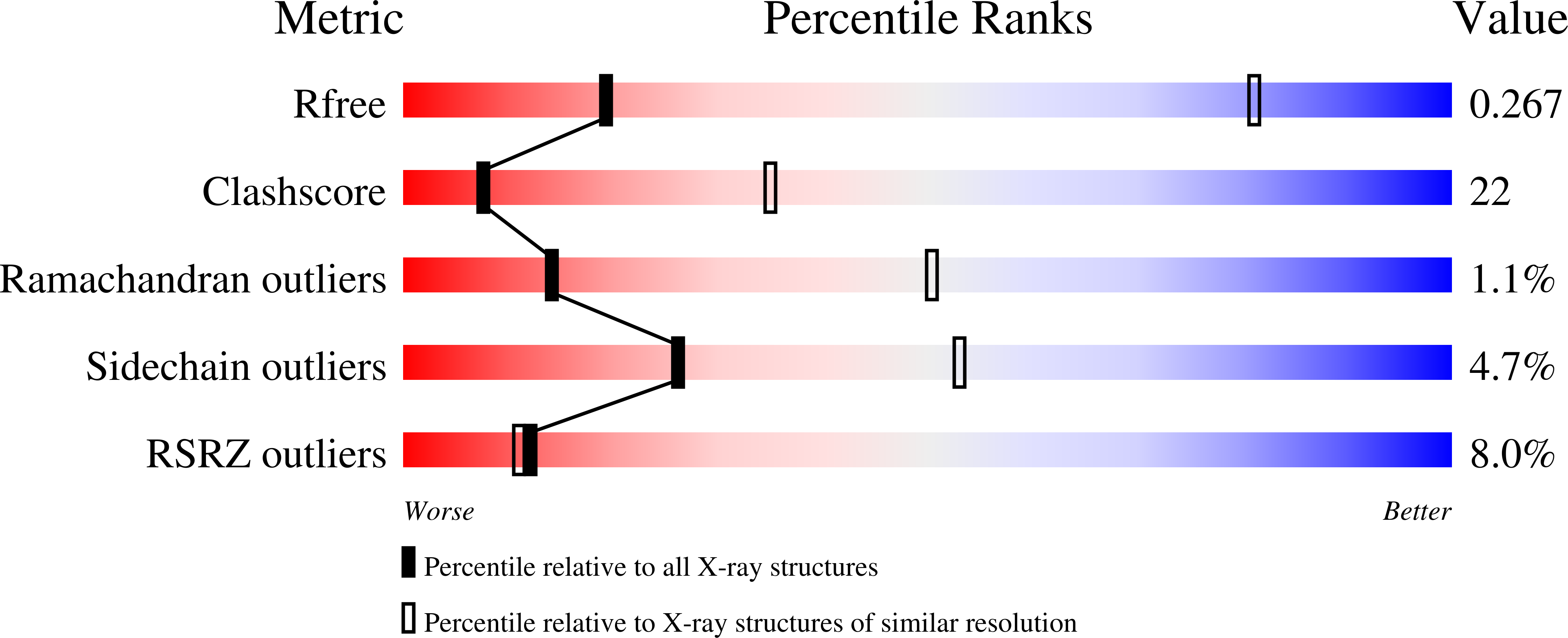
  • R-Value Free: 0.256 
  • R-Value Work: 0.224 
  • R-Value Observed: 0.225 

wwPDB Validation   3D Report Full Report


This is version 2.1 of the entry. See complete history


Literature

Crystal structures of Mycobacterium tuberculosis GlgE and complexes with non-covalent inhibitors.

Lindenberger, J.J.Kumar Veleti, S.Wilson, B.N.Sucheck, S.J.Ronning, D.R.

(2015) Sci Rep 5: 12830-12830

  • DOI: https://doi.org/10.1038/srep12830
  • Primary Citation of Related Structures:  
    4U2Y, 4U31, 4U33, 4U3C

  • PubMed Abstract: 

    GlgE is a bacterial maltosyltransferase that catalyzes the elongation of a cytosolic, branched α-glucan. In Mycobacterium tuberculosis (M. tb), inactivation of GlgE (Mtb GlgE) results in the rapid death of the organism due to a toxic accumulation of the maltosyl donor, maltose-1-phosphate (M1P), suggesting that GlgE is an intriguing target for inhibitor design. In this study, the crystal structures of the Mtb GlgE in a binary complex with maltose and a ternary complex with maltose and a maltosyl-acceptor molecule, maltohexaose, were solved to 3.3 Å and 4.0 Å, respectively. The maltohexaose structure reveals a dominant site for α-glucan binding. To obtain more detailed interactions between first generation, non-covalent inhibitors and GlgE, a variant Streptomyces coelicolor GlgEI (Sco GlgEI-V279S) was made to better emulate the Mtb GlgE M1P binding site. The structure of Sco GlgEI-V279S complexed with α-maltose-C-phosphonate (MCP), a non-hydrolyzable substrate analogue, was solved to 1.9 Å resolution, and the structure of Sco GlgEI-V279S complexed with 2,5-dideoxy-3-O-α-D-glucopyranosyl-2,5-imino-D-mannitol (DDGIM), an oxocarbenium mimic, was solved to 2.5 Å resolution. These structures detail important interactions that contribute to the inhibitory activity of these compounds, and provide information on future designs that may be exploited to improve upon these first generation GlgE inhibitors.


  • Organizational Affiliation

    Department of Chemistry and Biochemistry, The University of Toledo, 2801 W. Bancroft St. Ms602, Toledo, OH, United States.


Macromolecules
Find similar proteins by:  (by identity cutoff)  |  3D Structure
Entity ID: 1
MoleculeChains Sequence LengthOrganismDetailsImage
Alpha-1,4-glucan:maltose-1-phosphate maltosyltransferase
A, B, C, D, E
A, B, C, D, E, F
723Mycobacterium tuberculosis CDC1551Mutation(s): 0 
Gene Names: glgEMT1369
EC: 2.4.99.16
UniProt
Find proteins for P9WQ16 (Mycobacterium tuberculosis (strain CDC 1551 / Oshkosh))
Explore P9WQ16 
Go to UniProtKB:  P9WQ16
Entity Groups  
Sequence Clusters30% Identity50% Identity70% Identity90% Identity95% Identity100% Identity
UniProt GroupP9WQ16
Sequence Annotations
Expand
  • Reference Sequence
Oligosaccharides

Help

Entity ID: 2
MoleculeChains Length2D Diagram Glycosylation3D Interactions
alpha-D-glucopyranose-(1-4)-alpha-D-glucopyranose
G, I, K, M, O
G, I, K, M, O, Q
2N/A
Glycosylation Resources
GlyTouCan:  G07411ON
GlyCosmos:  G07411ON
Entity ID: 3
MoleculeChains Length2D Diagram Glycosylation3D Interactions
alpha-D-glucopyranose-(1-4)-alpha-D-glucopyranose-(1-4)-alpha-D-glucopyranose-(1-4)-alpha-D-glucopyranose-(1-4)-alpha-D-glucopyranose-(1-4)-alpha-D-glucopyranose
H, J, L, N, P
H, J, L, N, P, R
6N/A
Glycosylation Resources
GlyTouCan:  G91368RN
GlyCosmos:  G91368RN
GlyGen:  G91368RN
Experimental Data & Validation

Experimental Data

  • Method: X-RAY DIFFRACTION
  • Resolution: 3.98 Å
  • R-Value Free: 0.256 
  • R-Value Work: 0.224 
  • R-Value Observed: 0.225 
  • Space Group: C 1 2 1
Unit Cell:
Length ( Å )Angle ( ˚ )
a = 338.422α = 90
b = 239.418β = 134.9
c = 239.447γ = 90
Software Package:
Software NamePurpose
PHENIXrefinement
HKL-2000data collection
PDB_EXTRACTdata extraction
HKL-2000data reduction
HKL-2000data scaling

Structure Validation

View Full Validation Report


Entry History & Funding Information

Deposition Data


Funding OrganizationLocationGrant Number
National Institutes of Health/National Institute Of Allergy and Infectious Diseases (NIH/NIAID)United StatesAI105084

Revision History  (Full details and data files)

  • Version 1.0: 2015-07-22
    Type: Initial release
  • Version 1.1: 2015-08-12
    Changes: Database references
  • Version 1.2: 2015-08-19
    Changes: Database references
  • Version 1.3: 2017-09-20
    Changes: Author supporting evidence, Derived calculations, Refinement description
  • Version 1.4: 2019-12-11
    Changes: Author supporting evidence
  • Version 2.0: 2020-07-29
    Type: Remediation
    Reason: Carbohydrate remediation
    Changes: Advisory, Atomic model, Data collection, Derived calculations, Non-polymer description, Structure summary
  • Version 2.1: 2023-12-27
    Changes: Data collection, Database references, Structure summary