4RMJ

Human Sirt2 in complex with ADP ribose and nicotinamide


Experimental Data Snapshot

  • Method: X-RAY DIFFRACTION
  • Resolution: 1.87 Å
  • R-Value Free: 0.240 
  • R-Value Work: 0.210 
  • R-Value Observed: 0.211 

wwPDB Validation   3D Report Full Report


Ligand Structure Quality Assessment 


This is version 1.1 of the entry. See complete history


Literature

Selective Sirt2 inhibition by ligand-induced rearrangement of the active site.

Rumpf, T.Schiedel, M.Karaman, B.Roessler, C.North, B.J.Lehotzky, A.Olah, J.Ladwein, K.I.Schmidtkunz, K.Gajer, M.Pannek, M.Steegborn, C.Sinclair, D.A.Gerhardt, S.Ovadi, J.Schutkowski, M.Sippl, W.Einsle, O.Jung, M.

(2015) Nat Commun 6: 6263-6263

  • DOI: https://doi.org/10.1038/ncomms7263
  • Primary Citation of Related Structures:  
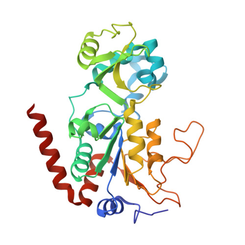
    4RMG, 4RMH, 4RMI, 4RMJ

  • PubMed Abstract: 

    Sirtuins are a highly conserved class of NAD(+)-dependent lysine deacylases. The human isotype Sirt2 has been implicated in the pathogenesis of cancer, inflammation and neurodegeneration, which makes the modulation of Sirt2 activity a promising strategy for pharmaceutical intervention. A rational basis for the development of optimized Sirt2 inhibitors is lacking so far. Here we present high-resolution structures of human Sirt2 in complex with highly selective drug-like inhibitors that show a unique inhibitory mechanism. Potency and the unprecedented Sirt2 selectivity are based on a ligand-induced structural rearrangement of the active site unveiling a yet-unexploited binding pocket. Application of the most potent Sirtuin-rearranging ligand, termed SirReal2, leads to tubulin hyperacetylation in HeLa cells and induces destabilization of the checkpoint protein BubR1, consistent with Sirt2 inhibition in vivo. Our structural insights into this unique mechanism of selective sirtuin inhibition provide the basis for further inhibitor development and selective tools for sirtuin biology.


  • Organizational Affiliation

    Institute of Pharmaceutical Sciences, Albert-Ludwigs-University Freiburg, Albertstraße 25, 79104 Freiburg im Breisgau, Germany.


Macromolecules
Find similar proteins by:  (by identity cutoff)  |  3D Structure
Entity ID: 1
MoleculeChains Sequence LengthOrganismDetailsImage
NAD-dependent protein deacetylase sirtuin-2
A, B
304Homo sapiensMutation(s): 0 
Gene Names: SIRT2SIR2LSIR2L2
EC: 3.5.1
UniProt & NIH Common Fund Data Resources
Find proteins for Q8IXJ6 (Homo sapiens)
Explore Q8IXJ6 
Go to UniProtKB:  Q8IXJ6
PHAROS:  Q8IXJ6
GTEx:  ENSG00000068903 
Entity Groups  
Sequence Clusters30% Identity50% Identity70% Identity90% Identity95% Identity100% Identity
UniProt GroupQ8IXJ6
Sequence Annotations
Expand
  • Reference Sequence
Small Molecules
Ligands 7 Unique
IDChains Name / Formula / InChI Key2D Diagram3D Interactions
AR6
Query on AR6

Download Ideal Coordinates CCD File 
C [auth A],
J [auth B]
[(2R,3S,4R,5R)-5-(6-AMINOPURIN-9-YL)-3,4-DIHYDROXY-OXOLAN-2-YL]METHYL [HYDROXY-[[(2R,3S,4R,5S)-3,4,5-TRIHYDROXYOXOLAN-2-YL]METHOXY]PHOSPHORYL] HYDROGEN PHOSPHATE
C15 H23 N5 O14 P2
SRNWOUGRCWSEMX-ZQSHOCFMSA-N
PGE
Query on PGE

Download Ideal Coordinates CCD File 
M [auth B]TRIETHYLENE GLYCOL
C6 H14 O4
ZIBGPFATKBEMQZ-UHFFFAOYSA-N
NCA
Query on NCA

Download Ideal Coordinates CCD File 
D [auth A]NICOTINAMIDE
C6 H6 N2 O
DFPAKSUCGFBDDF-UHFFFAOYSA-N
PEG
Query on PEG

Download Ideal Coordinates CCD File 
E [auth A]DI(HYDROXYETHYL)ETHER
C4 H10 O3
MTHSVFCYNBDYFN-UHFFFAOYSA-N
ZN
Query on ZN

Download Ideal Coordinates CCD File 
H [auth A],
I [auth B]
ZINC ION
Zn
PTFCDOFLOPIGGS-UHFFFAOYSA-N
EDO
Query on EDO

Download Ideal Coordinates CCD File 
F [auth A],
K [auth B],
L [auth B]
1,2-ETHANEDIOL
C2 H6 O2
LYCAIKOWRPUZTN-UHFFFAOYSA-N
NA
Query on NA

Download Ideal Coordinates CCD File 
G [auth A]SODIUM ION
Na
FKNQFGJONOIPTF-UHFFFAOYSA-N
Binding Affinity Annotations 
IDSourceBinding Affinity
NCA BindingDB:  4RMJ IC50: min: 1200, max: 1.01e+5 (nM) from 10 assay(s)
Experimental Data & Validation

Experimental Data

  • Method: X-RAY DIFFRACTION
  • Resolution: 1.87 Å
  • R-Value Free: 0.240 
  • R-Value Work: 0.210 
  • R-Value Observed: 0.211 
  • Space Group: P 21 21 21
Unit Cell:
Length ( Å )Angle ( ˚ )
a = 77.6α = 90
b = 77.96β = 90
c = 114.28γ = 90
Software Package:
Software NamePurpose
MOLREPphasing
REFMACrefinement
MOSFLMdata reduction
Aimlessdata scaling

Structure Validation

View Full Validation Report


Ligand Structure Quality Assessment 


Entry History 

Revision History  (Full details and data files)

  • Version 1.0: 2015-02-25
    Type: Initial release
  • Version 1.1: 2024-02-28
    Changes: Data collection, Database references, Derived calculations, Structure summary