


The crystal structure of Y333Q mutant pyridoxal-dependent decarboxylase from Sphaerobacter thermophilus DSM 20745
Wu, R., Clancy, S., Joachimiak, A., Midwest Center for Structural Genomics (MCSG)To be published.
Experimental Data Snapshot
Entity ID: 1 | |||||
|---|---|---|---|---|---|
| Molecule | Chains | Sequence Length | Organism | Details | Image |
| Pyridoxal-dependent decarboxylase | A [auth B], B [auth A], C, D | 486 | Sphaerobacter thermophilus DSM 20745 | Mutation(s): 1 Gene Names: Sthe_2364 EC: 4.1.1.15 | 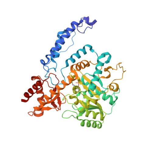 |
UniProt | |||||
Find proteins for D1C7D8 (Sphaerobacter thermophilus (strain DSM 20745 / S 6022)) Explore D1C7D8 Go to UniProtKB: D1C7D8 | |||||
Entity Groups | |||||
| Sequence Clusters | 30% Identity50% Identity70% Identity90% Identity95% Identity100% Identity | ||||
| UniProt Group | D1C7D8 | ||||
Sequence AnnotationsExpand | |||||
| |||||
| Ligands 3 Unique | |||||
|---|---|---|---|---|---|
| ID | Chains | Name / Formula / InChI Key | 2D Diagram | 3D Interactions | |
| PMP Query on PMP | E [auth B], M [auth C] | 4'-DEOXY-4'-AMINOPYRIDOXAL-5'-PHOSPHATE C8 H13 N2 O5 P ZMJGSOSNSPKHNH-UHFFFAOYSA-N |  | ||
| GOL Query on GOL | F [auth B] G [auth B] H [auth B] J [auth A] K [auth A] | GLYCEROL C3 H8 O3 PEDCQBHIVMGVHV-UHFFFAOYSA-N |  | ||
| CL Query on CL | I [auth B], N [auth D] | CHLORIDE ION Cl VEXZGXHMUGYJMC-UHFFFAOYSA-M |  | ||
| Modified Residues 1 Unique | |||||
|---|---|---|---|---|---|
| ID | Chains | Type | Formula | 2D Diagram | Parent |
| LLP Query on LLP | A [auth B], B [auth A], C, D | L-PEPTIDE LINKING | C14 H22 N3 O7 P |  | LYS |
| Length ( Å ) | Angle ( ˚ ) |
|---|---|
| a = 69.548 | α = 90 |
| b = 125.004 | β = 99.62 |
| c = 132.242 | γ = 90 |
| Software Name | Purpose |
|---|---|
| HKL-3000 | data collection |
| MLPHARE | phasing |
| PHENIX | refinement |
| HKL-3000 | data reduction |
| HKL-3000 | data scaling |
RCSB PDB (citation) is hosted by
RCSB PDB is a member of the

