4RAV

Crystal structure of scFvC4 in complex with the first 17 AA of huntingtin


Experimental Data Snapshot

  • Method: X-RAY DIFFRACTION
  • Resolution: 2.50 Å
  • R-Value Free: 0.226 
  • R-Value Work: 0.180 
  • R-Value Observed: 0.182 

wwPDB Validation   3D Report Full Report


This is version 1.0 of the entry. See complete history


Literature

Structure of a single-chain fv bound to the 17 N-terminal residues of huntingtin provides insights into pathogenic amyloid formation and suppression.

De Genst, E.Chirgadze, D.Y.Klein, F.A.Butler, D.C.Matak-Vinkovic, D.Trottier, Y.Huston, J.S.Messer, A.Dobson, C.M.

(2015) J Mol Biol 427: 2166-2178

  • DOI: https://doi.org/10.1016/j.jmb.2015.03.021
  • Primary Citation of Related Structures:  
    4RAV

  • PubMed Abstract: 

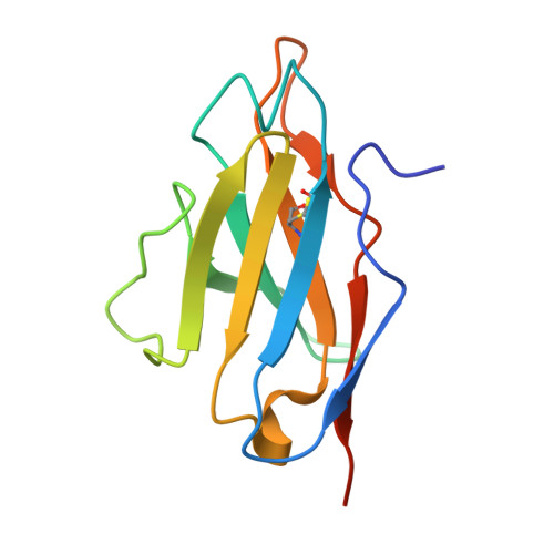
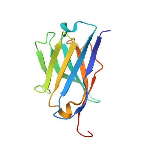
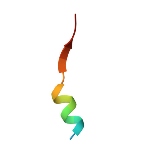
    Huntington's disease is triggered by misfolding of fragments of mutant forms of the huntingtin protein (mHTT) with aberrant polyglutamine expansions. The C4 single-chain Fv antibody (scFv) binds to the first 17 residues of huntingtin [HTT(1-17)] and generates substantial protection against multiple phenotypic pathologies in situ and in vivo. We show in this paper that C4 scFv inhibits amyloid formation by exon1 fragments of huntingtin in vitro and elucidate the structural basis for this inhibition and protection by determining the crystal structure of the complex of C4 scFv and HTT(1-17). The peptide binds with residues 3-11 forming an amphipathic helix that makes contact with the antibody fragment in such a way that the hydrophobic face of this helix is shielded from the solvent. Residues 12-17 of the peptide are in an extended conformation and interact with the same region of another C4 scFv:HTT(1-17) complex in the asymmetric unit, resulting in a β-sheet interface within a dimeric C4 scFv:HTT(1-17) complex. The nature of this scFv-peptide complex was further explored in solution by high-resolution NMR and physicochemical analysis of species in solution. The results provide insights into the manner in which C4 scFv inhibits the aggregation of HTT, and hence into its therapeutic potential, and suggests a structural basis for the initial interactions that underlie the formation of disease-associated amyloid fibrils by HTT.


  • Organizational Affiliation

    Department of Chemistry, University of Cambridge, Lensfield Road, Cambridge CB2 1EW, UK. Electronic address: ejjd2@cam.ac.uk.


Macromolecules
Find similar proteins by:  (by identity cutoff)  |  3D Structure
Entity ID: 1
MoleculeChains Sequence LengthOrganismDetailsImage
Single-chain Fv, VLA [auth B],
C [auth D]
117Homo sapiensMutation(s): 0 
Entity Groups  
Sequence Clusters30% Identity50% Identity70% Identity90% Identity95% Identity100% Identity
Sequence Annotations
Expand
  • Reference Sequence
Find similar proteins by:  (by identity cutoff)  |  3D Structure
Entity ID: 2
MoleculeChains Sequence LengthOrganismDetailsImage
single-chain Fv, VHB [auth A],
D [auth C]
126Homo sapiensMutation(s): 0 
Entity Groups  
Sequence Clusters30% Identity50% Identity70% Identity90% Identity95% Identity100% Identity
Sequence Annotations
Expand
  • Reference Sequence

Find similar proteins by:  Sequence   |   3D Structure  

Entity ID: 3
MoleculeChains Sequence LengthOrganismDetailsImage
Huntingtin
E, F
17Homo sapiensMutation(s): 0 
UniProt & NIH Common Fund Data Resources
Find proteins for P42858 (Homo sapiens)
Explore P42858 
Go to UniProtKB:  P42858
PHAROS:  P42858
GTEx:  ENSG00000197386 
Entity Groups  
Sequence Clusters30% Identity50% Identity70% Identity90% Identity95% Identity100% Identity
UniProt GroupP42858
Sequence Annotations
Expand
  • Reference Sequence
Experimental Data & Validation

Experimental Data

  • Method: X-RAY DIFFRACTION
  • Resolution: 2.50 Å
  • R-Value Free: 0.226 
  • R-Value Work: 0.180 
  • R-Value Observed: 0.182 
  • Space Group: C 1 2 1
Unit Cell:
Length ( Å )Angle ( ˚ )
a = 151.313α = 90
b = 35.933β = 120.72
c = 110.946γ = 90
Software Package:
Software NamePurpose
MxCuBEdata collection
PHENIXmodel building
PHENIXrefinement
XDSdata reduction
XSCALEdata scaling
PHENIXphasing

Structure Validation

View Full Validation Report


Entry History 

Deposition Data

Revision History  (Full details and data files)

  • Version 1.0: 2015-07-29
    Type: Initial release