4R6C

X-ray diffraction in temporally and spatially resolved biomolecular science: the X-ray crystal structure of hen egg white lysozyme cocrystallized with Ta6Br12 and then a crystal soaked in K2PtBr6


Experimental Data Snapshot

  • Method: X-RAY DIFFRACTION
  • Resolution: 1.70 Å
  • R-Value Free: 0.230 
  • R-Value Work: 0.180 
  • R-Value Observed: 0.183 

wwPDB Validation   3D Report Full Report


This is version 1.2 of the entry. See complete history


Literature

X-ray diffraction in temporally and spatially resolved biomolecular science.

Helliwell, J.R.Brink, A.Kaenket, S.Starkey, V.L.Tanley, S.W.

(2015) Faraday Discuss 177: 429-441

  • DOI: https://doi.org/10.1039/c4fd00166d
  • Primary Citation of Related Structures:  
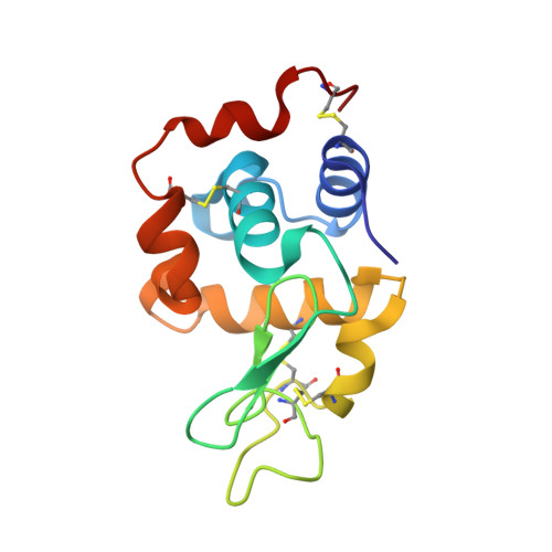
    4R6C

  • PubMed Abstract: 

    Time-resolved Laue protein crystallography at the European Synchrotron Radiation Facility (ESRF) opened up the field of sub-nanosecond protein crystal structure analyses. There are a limited number of such time-resolved studies in the literature. Why is this? The X-ray laser now gives us femtosecond (fs) duration pulses, typically 10 fs up to ∼50 fs. Their use is attractive for the fastest time-resolved protein crystallography studies. It has been proposed that single molecules could even be studied with the advantage of being able to measure X-ray diffraction from a 'crystal lattice free' single molecule, with or without temporal resolved structural changes. This is altogether very challenging R&D. So as to assist this effort we have undertaken studies of metal clusters that bind to proteins, both 'fresh' and after repeated X-ray irradiation to assess their X-ray-photo-dynamics, namely Ta6Br12, K2PtI6 and K2PtBr6 bound to a test protein, hen egg white lysozyme. These metal complexes have the major advantage of being very recognisable shapes (pseudo spherical or octahedral) and thereby offer a start to (probably very difficult) single molecule electron density map interpretations, both static and dynamic. A further approach is to investigate the X-ray laser beam diffraction strength of a well scattering nano-cluster; an example from nature being the iron containing ferritin. Electron crystallography and single particle electron microscopy imaging offers alternatives to X-ray structural studies; our structural studies of crustacyanin, a 320 kDa protein carotenoid complex, can be extended either by electron based techniques or with the X-ray laser representing a fascinating range of options. General outlook remarks concerning X-ray, electron and neutron macromolecular crystallography as well as 'NMR crystallography' conclude the article.


  • Organizational Affiliation

    School of Chemistry, University of Manchester M13 9PL, UK. john.helliwell@manchester.ac.uk.


Macromolecules
Find similar proteins by:  (by identity cutoff)  |  3D Structure
Entity ID: 1
MoleculeChains Sequence LengthOrganismDetailsImage
Lysozyme C129Gallus gallusMutation(s): 0 
EC: 3.2.1.17
UniProt
Find proteins for P00698 (Gallus gallus)
Explore P00698 
Go to UniProtKB:  P00698
Entity Groups  
Sequence Clusters30% Identity50% Identity70% Identity90% Identity95% Identity100% Identity
UniProt GroupP00698
Sequence Annotations
Expand
  • Reference Sequence
Small Molecules
Ligands 5 Unique
IDChains Name / Formula / InChI Key2D Diagram3D Interactions
TBR
Query on TBR

Download Ideal Coordinates CCD File 
B [auth A],
C [auth A]
HEXATANTALUM DODECABROMIDE
Br12 Ta6
YWYIQTPPCOBSGN-UHFFFAOYSA-M
6BP
Query on 6BP

Download Ideal Coordinates CCD File 
D [auth A],
E [auth A]
HEXABROMOPLATINATE(IV)
Br6 Pt
VOEHEFXBCNCSCK-UHFFFAOYSA-H
BR
Query on BR

Download Ideal Coordinates CCD File 
F [auth A]BROMIDE ION
Br
CPELXLSAUQHCOX-UHFFFAOYSA-M
CL
Query on CL

Download Ideal Coordinates CCD File 
G [auth A]
H [auth A]
I [auth A]
K [auth A]
L [auth A]
G [auth A],
H [auth A],
I [auth A],
K [auth A],
L [auth A],
M [auth A],
O [auth A],
P [auth A],
Q [auth A],
R [auth A],
S [auth A],
T [auth A]
CHLORIDE ION
Cl
VEXZGXHMUGYJMC-UHFFFAOYSA-M
NA
Query on NA

Download Ideal Coordinates CCD File 
J [auth A],
N [auth A]
SODIUM ION
Na
FKNQFGJONOIPTF-UHFFFAOYSA-N
Experimental Data & Validation

Experimental Data

  • Method: X-RAY DIFFRACTION
  • Resolution: 1.70 Å
  • R-Value Free: 0.230 
  • R-Value Work: 0.180 
  • R-Value Observed: 0.183 
  • Space Group: P 43 21 2
Unit Cell:
Length ( Å )Angle ( ˚ )
a = 79.07α = 90
b = 79.07β = 90
c = 37.235γ = 90
Software Package:
Software NamePurpose
PHASERphasing
REFMACrefinement
SAINTdata reduction

Structure Validation

View Full Validation Report


Entry History 

Deposition Data

Revision History  (Full details and data files)

  • Version 1.0: 2015-02-04
    Type: Initial release
  • Version 1.1: 2015-04-29
    Changes: Database references
  • Version 1.2: 2018-03-07
    Changes: Data collection