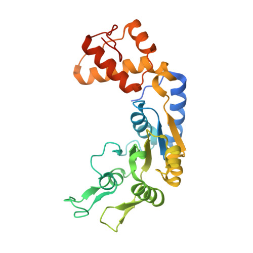
4QHT

Crystal structure of AAA+/ sigma 54 activator domain of the flagellar regulatory protein FlrC from Vibrio cholerae in ATP analog bound state


Experimental Data Snapshot

  • Method: X-RAY DIFFRACTION
  • Resolution: 2.56 Å
  • R-Value Free: 0.247 
  • R-Value Work: 0.182 
  • R-Value Observed: 0.186 

wwPDB Validation   3D Report Full Report


Ligand Structure Quality Assessment 


This is version 1.2 of the entry. See complete history


Literature

Unique ATPase site architecture triggers cis-mediated synchronized ATP binding in heptameric AAA+-ATPase domain of flagellar regulatory protein FlrC

Dey, S.Biswas, M.Sen, U.Dasgupta, J.

(2015) J Biol Chem 290: 8734-8747

  • DOI: https://doi.org/10.1074/jbc.M114.611434
  • Primary Citation of Related Structures:  
    4QHS, 4QHT

  • PubMed Abstract: 

    Bacterial enhancer-binding proteins (bEBPs) oligomerize through AAA(+) domains and use ATP hydrolysis-driven energy to isomerize the RNA polymerase-σ(54) complex during transcriptional initiation. Here, we describe the first structure of the central AAA(+) domain of the flagellar regulatory protein FlrC (FlrC(C)), a bEBP that controls flagellar synthesis in Vibrio cholerae. Our results showed that FlrC(C) forms heptamer both in nucleotide (Nt)-free and -bound states without ATP-dependent subunit remodeling. Unlike the bEBPs such as NtrC1 or PspF, a novel cis-mediated "all or none" ATP binding occurs in the heptameric FlrC(C), because constriction at the ATPase site, caused by loop L3 and helix α7, restricts the proximity of the trans-protomer required for Nt binding. A unique "closed to open" movement of Walker A, assisted by trans-acting "Glu switch" Glu-286, facilitates ATP binding and hydrolysis. Fluorescence quenching and ATPase assays on FlrC(C) and mutants revealed that although Arg-349 of sensor II, positioned by trans-acting Glu-286 and Tyr-290, acts as a key residue to bind and hydrolyze ATP, Arg-319 of α7 anchors ribose and controls the rate of ATP hydrolysis by retarding the expulsion of ADP. Heptameric state of FlrC(C) is restored in solution even with the transition state mimicking ADP·AlF3. Structural results and pulldown assays indicated that L3 renders an in-built geometry to L1 and L2 causing σ(54)-FlrC(C) interaction independent of Nt binding. Collectively, our results underscore a novel mechanism of ATP binding and σ(54) interaction that strives to understand the transcriptional mechanism of the bEBPs, which probably interact directly with the RNA polymerase-σ(54) complex without DNA looping.


  • Organizational Affiliation

    From the Department of Biotechnology, St. Xavier's College, 30 Park Street, Kolkata 700016 and.


Macromolecules
Find similar proteins by:  (by identity cutoff)  |  3D Structure
Entity ID: 1
MoleculeChains Sequence LengthOrganismDetailsImage
Flagellar regulatory protein C
A, B, C, D, E
A, B, C, D, E, F, G
267Vibrio cholerae O395Mutation(s): 0 
Gene Names: flrC
UniProt
Find proteins for A0A0H3AHP1 (Vibrio cholerae serotype O1 (strain ATCC 39541 / Classical Ogawa 395 / O395))
Explore A0A0H3AHP1 
Go to UniProtKB:  A0A0H3AHP1
Entity Groups  
Sequence Clusters30% Identity50% Identity70% Identity90% Identity95% Identity100% Identity
UniProt GroupA0A0H3AHP1
Sequence Annotations
Expand
  • Reference Sequence
Small Molecules
Ligands 3 Unique
IDChains Name / Formula / InChI Key2D Diagram3D Interactions
ANP
Query on ANP

Download Ideal Coordinates CCD File 
DA [auth G]
H [auth A]
L [auth B]
O [auth C]
R [auth D]
DA [auth G],
H [auth A],
L [auth B],
O [auth C],
R [auth D],
V [auth E],
Z [auth F]
PHOSPHOAMINOPHOSPHONIC ACID-ADENYLATE ESTER
C10 H17 N6 O12 P3
PVKSNHVPLWYQGJ-KQYNXXCUSA-N
EDO
Query on EDO

Download Ideal Coordinates CCD File 
BA [auth F]
CA [auth F]
FA [auth G]
GA [auth G]
J [auth A]
BA [auth F],
CA [auth F],
FA [auth G],
GA [auth G],
J [auth A],
K [auth A],
N [auth B],
Q [auth C],
T [auth D],
U [auth D],
X [auth E],
Y [auth E]
1,2-ETHANEDIOL
C2 H6 O2
LYCAIKOWRPUZTN-UHFFFAOYSA-N
MG
Query on MG

Download Ideal Coordinates CCD File 
AA [auth F]
EA [auth G]
I [auth A]
M [auth B]
P [auth C]
AA [auth F],
EA [auth G],
I [auth A],
M [auth B],
P [auth C],
S [auth D],
W [auth E]
MAGNESIUM ION
Mg
JLVVSXFLKOJNIY-UHFFFAOYSA-N
Experimental Data & Validation

Experimental Data

  • Method: X-RAY DIFFRACTION
  • Resolution: 2.56 Å
  • R-Value Free: 0.247 
  • R-Value Work: 0.182 
  • R-Value Observed: 0.186 
  • Space Group: P 21 21 21
Unit Cell:
Length ( Å )Angle ( ˚ )
a = 80.959α = 90
b = 152.56β = 90
c = 193.138γ = 90
Software Package:
Software NamePurpose
HKL-2000data collection
PHASERphasing
PHENIXrefinement
HKL-2000data reduction
HKL-2000data scaling

Structure Validation

View Full Validation Report


Ligand Structure Quality Assessment 


Entry History 

Deposition Data

Revision History  (Full details and data files)

  • Version 1.0: 2014-07-16
    Type: Initial release
  • Version 1.1: 2015-10-14
    Changes: Database references
  • Version 1.2: 2024-03-20
    Changes: Data collection, Database references, Derived calculations