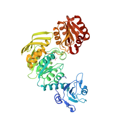
ATP-binding mode including a carbamoylated lysine and two Mg(2+) ions, and substrate-binding mode in Acinetobacter baumannii MurF
Cha, S.S., An, Y.J., Jeong, C.S., Yu, J.H., Chung, K.M.(2014) Biochem Biophys Res Commun 450: 1045-1050
- PubMed: 24978312
- DOI: https://doi.org/10.1016/j.bbrc.2014.06.108
- Primary Citation of Related Structures:
4QDI, 4QF5 - PubMed Abstract:
MurF adds d-Ala-d-Ala dipeptide to UDP-N-acetylmuramyl-l-Ala-γ-d-Glu-m-DAP (or l-Lys) in an ATP-dependent manner, which is the last step in the biosynthesis of monomeric precursor of peptidoglycan. Here we report crystal structures of two MurF-ATP complexes: the MurF-ATP complex and the MurF-ATP-UDP complex. The ATP-binding mode revealed by the crystal structure of the MurF-ATP complex confirms the previous biochemical demonstration that a carbamoylated lysine and two Mg(2+) ions are required for enzyme activity of MurF. The UDP-MurF interactions observed in the crystal structure of the MurF-ATP-UDP complex depict the characteristic substrate-binding mode of MurF. The emergence and dissemination of multidrug-resistant Acinetobacter baumannii strains are great threats to public health. Therefore, the structural information on A. baumannii MurF as a validated target for drug discovery will provide a framework to develop antibacterial agents against multidrug-resistant A. baumannii infections as well as to understand the reaction mechanism of MurF.
Organizational Affiliation:
Marine Biotechnology Research Division, Korea Institute of Ocean Science and Technology, Ansan 426-744, Republic of Korea; Department of Convergence Study on the Ocean Science and Technology, Ocean Science and Technology School, Pusan 606-791, Republic of Korea; Department of Marine Biotechnology, Korea University of Science and Technology, Daejeon 305-333, Republic of Korea. Electronic address: chajung@kiost.ac.