4Q0P

Crystal structure of Acinetobacter sp. DL28 L-ribose isomerase in complex with L-ribose


Experimental Data Snapshot

  • Method: X-RAY DIFFRACTION
  • Resolution: 1.93 Å
  • R-Value Free: 0.242 
  • R-Value Work: 0.208 
  • R-Value Observed: 0.208 

wwPDB Validation   3D Report Full Report


Ligand Structure Quality Assessment 


This is version 2.2 of the entry. See complete history


Literature

X-ray structure of a novel L-ribose isomerase acting on a non-natural sugar L-ribose as its ideal substrate.

Yoshida, H.Yoshihara, A.Teraoka, M.Terami, Y.Takata, G.Izumori, K.Kamitori, S.

(2014) FEBS J 281: 3150-3164

  • DOI: https://doi.org/10.1111/febs.12850
  • Primary Citation of Related Structures:  
    4Q0P, 4Q0S

  • PubMed Abstract: 

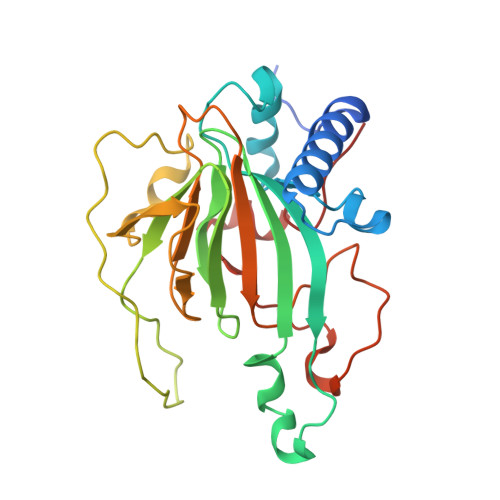
    l-Ribose, a pentose, is not known to exist in nature. Although organisms typically do not have a metabolic pathway that uses l-ribose as a carbon source, prokaryotes use various sugars as carbon sources for survival. Acinetobacter sp. DL-28 has been shown to express the novel enzyme, l-ribose isomerase (AcL-RbI), which catalyzes reversible isomerization between l-ribose and l-ribulose. AcL-RbI showed the highest activity to l-ribose, followed by d-lyxose with 47% activity, and had no significant amino acid sequence similarity to structure-known proteins, except for weak homology with the d-lyxose isomerases from Escherichia coli O157 : H7 (18%) and Bacillus subtilis strain (19%). Thus, AcL-RbI is expected to have the unique three-dimensional structure to recognize l-ribose as its ideal substrate. The X-ray structures of AcL-RbI in complexes with substrates were determined. AcL-RbI had a cupin-type β-barrel structure, and the catalytic site was found between two large β-sheets with a bound metal ion. The catalytic site structures clearly showed that AcL-RbI adopted a cis-enediol intermediate mechanism for the isomerization reaction using two glutamate residues (Glu113 and Glu204) as acid/base catalysts. In its crystal form, AcL-RbI formed a unique homotetramer with many substrate sub-binding sites, which likely facilitated capture of the substrate.


  • Organizational Affiliation

    Life Science Research Center and Faculty of Medicine, Kagawa University, Japan.


Macromolecules
Find similar proteins by:  (by identity cutoff)  |  3D Structure
Entity ID: 1
MoleculeChains Sequence LengthOrganismDetailsImage
L-Ribose isomerase260Acinetobacter sp. DL-28Mutation(s): 0 
Gene Names: L-RI
EC: 5.3.1
UniProt
Find proteins for Q93UQ5 (Acinetobacter sp. DL-28)
Explore Q93UQ5 
Go to UniProtKB:  Q93UQ5
Entity Groups  
Sequence Clusters30% Identity50% Identity70% Identity90% Identity95% Identity100% Identity
UniProt GroupQ93UQ5
Sequence Annotations
Expand
  • Reference Sequence
Experimental Data & Validation

Experimental Data

  • Method: X-RAY DIFFRACTION
  • Resolution: 1.93 Å
  • R-Value Free: 0.242 
  • R-Value Work: 0.208 
  • R-Value Observed: 0.208 
  • Space Group: F 2 2 2
Unit Cell:
Length ( Å )Angle ( ˚ )
a = 95.38α = 90
b = 107.93β = 90
c = 119.41γ = 90
Software Package:
Software NamePurpose
CrystalCleardata collection
MOLREPphasing
CNSrefinement
CrystalCleardata reduction
CrystalCleardata scaling

Structure Validation

View Full Validation Report


Ligand Structure Quality Assessment 


Entry History 

Deposition Data

Revision History  (Full details and data files)

  • Version 1.0: 2014-05-28
    Type: Initial release
  • Version 1.1: 2014-06-11
    Changes: Database references
  • Version 2.0: 2020-07-29
    Type: Remediation
    Reason: Carbohydrate remediation
    Changes: Atomic model, Data collection, Database references, Derived calculations, Structure summary
  • Version 2.1: 2022-08-24
    Changes: Database references, Structure summary
  • Version 2.2: 2024-04-03
    Changes: Data collection, Refinement description