4PZD

Crystal structure of (S)-3-hydroxybutyryl-CoA dehydrogenase PaaH1 in complex with NAD+


Experimental Data Snapshot

  • Method: X-RAY DIFFRACTION
  • Resolution: 2.61 Å
  • R-Value Free: 0.269 
  • R-Value Work: 0.199 
  • R-Value Observed: 0.202 

wwPDB Validation   3D Report Full Report


Ligand Structure Quality Assessment 


This is version 1.2 of the entry. See complete history


Literature

Crystal structure and biochemical properties of the (S)-3-hydroxybutyryl-CoA dehydrogenase PaaH1 from Ralstonia eutropha

Kim, J.Chang, J.H.Kim, K.J.

(2014) Biochem Biophys Res Commun 448: 163-168

  • DOI: https://doi.org/10.1016/j.bbrc.2014.04.101
  • Primary Citation of Related Structures:  
    4PZC, 4PZD, 4PZE

  • PubMed Abstract: 

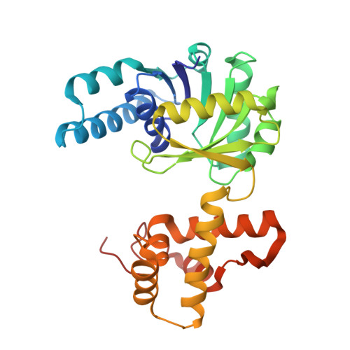
    3-Hydroxybutyryl-CoA dehydrogenase is an enzyme involved in the synthesis of the biofuel n-butanol by converting acetoacetyl-CoA to 3-hydroxybutyryl-CoA. To investigate the molecular mechanism of n-butanol biosynthesis, we determined crystal structures of the Ralstonia eutropha-derived 3-hydroxybutyryl-CoA dehydrogenase (RePaaH1) in complex with either its cofactor NAD(+) or its substrate acetoacetyl-CoA. While the biologically active structure is dimeric, the monomer of RePaaH1 comprises two separated domains with an N-terminal Rossmann fold and a C-terminal helical bundle for dimerization. In this study, we show that the cofactor-binding site is located on the Rossmann fold and is surrounded by five loops and one helix. The binding mode of the acetoacetyl-CoA substrate was found to be that the adenosine diphosphate moiety is not highly stabilized compared with the remainder of the molecule. Residues involved in catalysis and substrate binding were further confirmed by site-directed mutagenesis experiments, and kinetic properties of RePaaH1were examined as well. Our findings contribute to the understanding of 3-hydroxybutyryl-CoA dehydrogenase catalysis, and will be useful in enhancing the efficiency of n-butanol biosynthesis by structure based protein engineering.


  • Organizational Affiliation

    School of Life Sciences, KNU Creative BioResearch Group (BK21 Plus Program), Kyungpook National University, Daehak-ro 80, Buk-ku, Daegu 702-701, Republic of Korea.


Macromolecules
Find similar proteins by:  (by identity cutoff)  |  3D Structure
Entity ID: 1
MoleculeChains Sequence LengthOrganismDetailsImage
3-Hydroxyacyl-CoA dehydrogenase
A, B, C, D, E
A, B, C, D, E, F, G, H, I
284Cupriavidus necator H16Mutation(s): 0 
Gene Names: paaH1H16_A0282
EC: 1.1.1.35
UniProt
Find proteins for Q0KEY8 (Cupriavidus necator (strain ATCC 17699 / DSM 428 / KCTC 22496 / NCIMB 10442 / H16 / Stanier 337))
Explore Q0KEY8 
Go to UniProtKB:  Q0KEY8
Entity Groups  
Sequence Clusters30% Identity50% Identity70% Identity90% Identity95% Identity100% Identity
UniProt GroupQ0KEY8
Sequence Annotations
Expand
  • Reference Sequence
Experimental Data & Validation

Experimental Data

  • Method: X-RAY DIFFRACTION
  • Resolution: 2.61 Å
  • R-Value Free: 0.269 
  • R-Value Work: 0.199 
  • R-Value Observed: 0.202 
  • Space Group: C 1 2 1
Unit Cell:
Length ( Å )Angle ( ˚ )
a = 235.076α = 90
b = 135.586β = 90.09
c = 97.445γ = 90
Software Package:
Software NamePurpose
HKL-2000data collection
MLPHAREphasing
REFMACrefinement
HKL-2000data reduction
HKL-2000data scaling

Structure Validation

View Full Validation Report


Ligand Structure Quality Assessment 


Entry History 

Deposition Data

Revision History  (Full details and data files)

  • Version 1.0: 2015-02-11
    Type: Initial release
  • Version 1.1: 2015-02-18
    Changes: Derived calculations
  • Version 1.2: 2024-03-20
    Changes: Data collection, Database references, Derived calculations