4PXZ

Crystal structure of P2Y12 receptor in complex with 2MeSADP


Experimental Data Snapshot

  • Method: X-RAY DIFFRACTION
  • Resolution: 2.50 Å
  • R-Value Free: 0.230 
  • R-Value Work: 0.201 
  • R-Value Observed: 0.202 

wwPDB Validation   3D Report Full Report


Ligand Structure Quality Assessment 


This is version 1.5 of the entry. See complete history


Literature

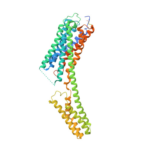
Agonist-bound structure of the human P2Y12 receptor

Zhang, J.Zhang, K.Gao, Z.G.Paoletta, S.Zhang, D.Han, G.W.Li, T.Ma, L.Zhang, W.Muller, C.E.Yang, H.Jiang, H.Cherezov, V.Katritch, V.Jacobson, K.A.Stevens, R.C.Wu, B.Zhao, Q.

(2014) Nature 509: 119-122

  • DOI: https://doi.org/10.1038/nature13288
  • Primary Citation of Related Structures:  
    4PXZ, 4PY0

  • PubMed Abstract: 

    The P2Y12 receptor (P2Y12R), one of eight members of the P2YR family expressed in humans, is one of the most prominent clinical drug targets for inhibition of platelet aggregation. Although mutagenesis and modelling studies of the P2Y12R provided useful insights into ligand binding, the agonist and antagonist recognition and function at the P2Y12R remain poorly understood at the molecular level. Here we report the structures of the human P2Y12R in complex with the full agonist 2-methylthio-adenosine-5'-diphosphate (2MeSADP, a close analogue of endogenous agonist ADP) at 2.5 Å resolution, and the corresponding ATP derivative 2-methylthio-adenosine-5'-triphosphate (2MeSATP) at 3.1 Å resolution. These structures, together with the structure of the P2Y12R with antagonist ethyl 6-(4-((benzylsulfonyl)carbamoyl)piperidin-1-yl)-5-cyano-2-methylnicotinate (AZD1283), reveal striking conformational changes between nucleotide and non-nucleotide ligand complexes in the extracellular regions. Further analysis of these changes provides insight into a distinct ligand binding landscape in the δ-group of class A G-protein-coupled receptors (GPCRs). Agonist and non-nucleotide antagonist adopt different orientations in the P2Y12R, with only partially overlapped binding pockets. The agonist-bound P2Y12R structure answers long-standing questions surrounding P2Y12R-agonist recognition, and reveals interactions with several residues that had not been reported to be involved in agonist binding. As a first example, to our knowledge, of a GPCR in which agonist access to the binding pocket requires large-scale rearrangements in the highly malleable extracellular region, the structural and docking studies will therefore provide invaluable insight into the pharmacology and mechanisms of action of agonists and different classes of antagonists for the P2Y12R and potentially for other closely related P2YRs.


  • Organizational Affiliation

    1] CAS Key Laboratory of Receptor Research, Shanghai Institute of Materia Medica, Chinese Academy of Sciences, 555 Zuchongzhi Road, Pudong, Shanghai 201203, China [2].


Macromolecules
Find similar proteins by:  (by identity cutoff)  |  3D Structure
Entity ID: 1
MoleculeChains Sequence LengthOrganismDetailsImage
P2Y purinoceptor 12, Soluble cytochrome b562466Homo sapiensEscherichia coliMutation(s): 4 
Gene Names: HORK3P2RY12P2Y12cybC
Membrane Entity: Yes 
UniProt & NIH Common Fund Data Resources
Find proteins for P0ABE7 (Escherichia coli)
Explore P0ABE7 
Go to UniProtKB:  P0ABE7
Find proteins for Q9H244 (Homo sapiens)
Explore Q9H244 
Go to UniProtKB:  Q9H244
PHAROS:  Q9H244
GTEx:  ENSG00000169313 
Entity Groups  
Sequence Clusters30% Identity50% Identity70% Identity90% Identity95% Identity100% Identity
UniProt GroupsQ9H244P0ABE7
Sequence Annotations
Expand
  • Reference Sequence
Small Molecules
Ligands 4 Unique
IDChains Name / Formula / InChI Key2D Diagram3D Interactions
6AD
Query on 6AD

Download Ideal Coordinates CCD File 
B [auth A]2-(methylsulfanyl)adenosine 5'-(trihydrogen diphosphate)
C11 H17 N5 O10 P2 S
WLMZTKAZJUWXCB-KQYNXXCUSA-N
CLR
Query on CLR

Download Ideal Coordinates CCD File 
C [auth A]CHOLESTEROL
C27 H46 O
HVYWMOMLDIMFJA-DPAQBDIFSA-N
OLC
Query on OLC

Download Ideal Coordinates CCD File 
D [auth A],
E [auth A],
F [auth A]
(2R)-2,3-dihydroxypropyl (9Z)-octadec-9-enoate
C21 H40 O4
RZRNAYUHWVFMIP-GDCKJWNLSA-N
PEG
Query on PEG

Download Ideal Coordinates CCD File 
G [auth A]DI(HYDROXYETHYL)ETHER
C4 H10 O3
MTHSVFCYNBDYFN-UHFFFAOYSA-N
Experimental Data & Validation

Experimental Data

  • Method: X-RAY DIFFRACTION
  • Resolution: 2.50 Å
  • R-Value Free: 0.230 
  • R-Value Work: 0.201 
  • R-Value Observed: 0.202 
  • Space Group: C 2 2 21
Unit Cell:
Length ( Å )Angle ( ˚ )
a = 65.11α = 90
b = 104.17β = 90
c = 169.43γ = 90
Software Package:
Software NamePurpose
Blu-Icedata collection
PHASERphasing
BUSTERrefinement
XDSdata reduction
XDSdata scaling

Structure Validation

View Full Validation Report


Ligand Structure Quality Assessment 


Entry History 

Revision History  (Full details and data files)

  • Version 1.0: 2014-04-30
    Type: Initial release
  • Version 1.1: 2014-05-28
    Changes: Database references
  • Version 1.2: 2014-09-03
    Changes: Derived calculations
  • Version 1.3: 2017-08-16
    Changes: Refinement description, Source and taxonomy
  • Version 1.4: 2017-11-22
    Changes: Refinement description
  • Version 1.5: 2023-11-08
    Changes: Data collection, Database references, Derived calculations, Refinement description