4PWA

Crystal structure of the c-type cytochrome SorU from Sinorhizobium meliloti


Experimental Data Snapshot

  • Method: X-RAY DIFFRACTION
  • Resolution: 2.19 Å
  • R-Value Free: 0.241 
  • R-Value Work: 0.185 
  • R-Value Observed: 0.188 

wwPDB Validation   3D Report Full Report


Ligand Structure Quality Assessment 


This is version 1.3 of the entry. See complete history


Literature

Structural basis of interprotein electron transfer in bacterial sulfite oxidation.

McGrath, A.P.Laming, E.L.Casas Garcia, G.P.Kvansakul, M.Guss, J.M.Trewhella, J.Calmes, B.Bernhardt, P.V.Hanson, G.R.Kappler, U.Maher, M.J.

(2015) Elife 4: e09066-e09066

  • DOI: https://doi.org/10.7554/eLife.09066
  • Primary Citation of Related Structures:  
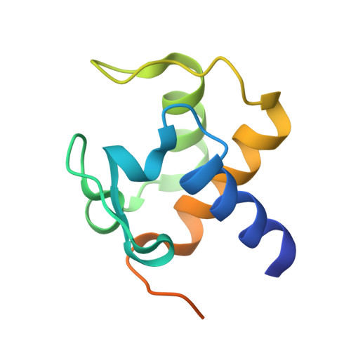
    4PW3, 4PW9, 4PWA

  • PubMed Abstract: 

    Interprotein electron transfer underpins the essential processes of life and relies on the formation of specific, yet transient protein-protein interactions. In biological systems, the detoxification of sulfite is catalyzed by the sulfite-oxidizing enzymes (SOEs), which interact with an electron acceptor for catalytic turnover. Here, we report the structural and functional analyses of the SOE SorT from Sinorhizobium meliloti and its cognate electron acceptor SorU. Kinetic and thermodynamic analyses of the SorT/SorU interaction show the complex is dynamic in solution, and that the proteins interact with Kd = 13.5 ± 0.8 μM. The crystal structures of the oxidized SorT and SorU, both in isolation and in complex, reveal the interface to be remarkably electrostatic, with an unusually large number of direct hydrogen bonding interactions. The assembly of the complex is accompanied by an adjustment in the structure of SorU, and conformational sampling provides a mechanism for dissociation of the SorT/SorU assembly.


  • Organizational Affiliation

    Structural Biology Program, Centenary Institute, Sydney, Australia.


Macromolecules
Find similar proteins by:  (by identity cutoff)  |  3D Structure
Entity ID: 1
MoleculeChains Sequence LengthOrganismDetailsImage
Putative cytochrome C
A, B, C, D
106Sinorhizobium meliloti 1021Mutation(s): 0 
Gene Names: R02831SMc04048
UniProt
Find proteins for Q92M25 (Rhizobium meliloti (strain 1021))
Explore Q92M25 
Go to UniProtKB:  Q92M25
Entity Groups  
Sequence Clusters30% Identity50% Identity70% Identity90% Identity95% Identity100% Identity
UniProt GroupQ92M25
Sequence Annotations
Expand
  • Reference Sequence
Experimental Data & Validation

Experimental Data

  • Method: X-RAY DIFFRACTION
  • Resolution: 2.19 Å
  • R-Value Free: 0.241 
  • R-Value Work: 0.185 
  • R-Value Observed: 0.188 
  • Space Group: F 2 2 2
Unit Cell:
Length ( Å )Angle ( ˚ )
a = 70.949α = 90
b = 129.327β = 90
c = 197.059γ = 90
Software Package:
Software NamePurpose
PHASERphasing
REFMACrefinement
HKL-2000data reduction
SCALEPACKdata scaling

Structure Validation

View Full Validation Report


Ligand Structure Quality Assessment 


Entry History 

Deposition Data

Revision History  (Full details and data files)

  • Version 1.0: 2015-06-03
    Type: Initial release
  • Version 1.1: 2016-02-17
    Changes: Database references
  • Version 1.2: 2017-02-15
    Changes: Database references
  • Version 1.3: 2023-09-20
    Changes: Data collection, Database references, Derived calculations, Refinement description