4PNO

Escherichia coli Hfq-RNA complex at 0.97 A Resolution


Experimental Data Snapshot

  • Method: X-RAY DIFFRACTION
  • Resolution: 0.97 Å
  • R-Value Free: 0.164 
  • R-Value Work: 0.142 
  • R-Value Observed: 0.144 

wwPDB Validation   3D Report Full Report


Ligand Structure Quality Assessment 


This is version 1.3 of the entry. See complete history


Literature

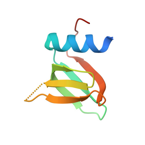
Structure of an Escherichia coli Hfq:RNA complex at 0.97 angstrom resolution.

Schulz, E.C.Barabas, O.

(2014) Acta Crystallogr F Struct Biol Commun 70: 1492-1497

  • DOI: https://doi.org/10.1107/S2053230X14020044
  • Primary Citation of Related Structures:  
    4PNO

  • PubMed Abstract: 

    In bacteria, small RNAs (sRNAs) silence or activate target genes through base pairing with the mRNA, thereby modulating its translation. A central player in this process is the RNA chaperone Hfq, which facilitates the annealing of sRNAs with their target mRNAs. Hfq has two RNA-binding surfaces that recognize A-rich and U-rich sequences, and is believed to bind an sRNA-mRNA pair simultaneously. However, how Hfq promotes annealing remains unclear. Here, the crystal structure of Escherichia coli Hfq is presented in complex with U6-RNA bound to its proximal binding site at 0.97 Å resolution, revealing the Hfq-RNA interaction in exceptional detail.


  • Organizational Affiliation

    Structural and Computational Biology Unit, European Molecular Biology Laboratory, Meyerhofstrasse 1, 69117 Heidelberg, Germany.


Macromolecules
Find similar proteins by:  (by identity cutoff)  |  3D Structure
Entity ID: 1
MoleculeChains Sequence LengthOrganismDetailsImage
RNA-binding protein Hfq72Escherichia coliMutation(s): 0 
Gene Names: hfqBN17_41491BU34_16545ECs5148LF82_0993
UniProt
Find proteins for C1IFD2 (Escherichia coli)
Explore C1IFD2 
Go to UniProtKB:  C1IFD2
Entity Groups  
Sequence Clusters30% Identity50% Identity70% Identity90% Identity95% Identity100% Identity
UniProt GroupC1IFD2
Sequence Annotations
Expand
  • Reference Sequence
Small Molecules
Ligands 1 Unique
IDChains Name / Formula / InChI Key2D Diagram3D Interactions
U5P
Query on U5P

Download Ideal Coordinates CCD File 
B [auth A]URIDINE-5'-MONOPHOSPHATE
C9 H13 N2 O9 P
DJJCXFVJDGTHFX-XVFCMESISA-N
Experimental Data & Validation

Experimental Data

  • Method: X-RAY DIFFRACTION
  • Resolution: 0.97 Å
  • R-Value Free: 0.164 
  • R-Value Work: 0.142 
  • R-Value Observed: 0.144 
  • Space Group: P 6
Unit Cell:
Length ( Å )Angle ( ˚ )
a = 61.11α = 90
b = 61.11β = 90
c = 27.86γ = 120
Software Package:
Software NamePurpose
PHENIXrefinement

Structure Validation

View Full Validation Report


Ligand Structure Quality Assessment 


Entry History & Funding Information

Deposition Data


Funding OrganizationLocationGrant Number
EU FP7 - Marie Curie IEFGermanyPIEF-GA-2011-301489

Revision History  (Full details and data files)

  • Version 1.0: 2014-11-19
    Type: Initial release
  • Version 1.1: 2014-11-26
    Changes: Data collection
  • Version 1.2: 2018-03-07
    Changes: Advisory, Data collection, Derived calculations, Other, Source and taxonomy
  • Version 1.3: 2023-09-27
    Changes: Advisory, Data collection, Database references, Refinement description