4PJR

Crystal structure of designed cPPR-NRE protein


Experimental Data Snapshot

  • Method: X-RAY DIFFRACTION
  • Resolution: 2.00 Å
  • R-Value Free: 0.222 
  • R-Value Work: 0.185 
  • R-Value Observed: 0.189 

wwPDB Validation   3D Report Full Report


This is version 1.5 of the entry. See complete history


Literature

An artificial PPR scaffold for programmable RNA recognition.

Coquille, S.Filipovska, A.Chia, T.Rajappa, L.Lingford, J.P.Razif, M.F.Thore, S.Rackham, O.

(2014) Nat Commun 5: 5729-5729

  • DOI: https://doi.org/10.1038/ncomms6729
  • Primary Citation of Related Structures:  
    4PJQ, 4PJR, 4PJS, 4WN4, 4WSL

  • PubMed Abstract: 

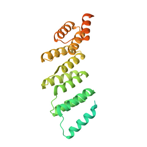
    Pentatricopeptide repeat (PPR) proteins control diverse aspects of RNA metabolism in eukaryotic cells. Although recent computational and structural studies have provided insights into RNA recognition by PPR proteins, their highly insoluble nature and inconsistencies between predicted and observed modes of RNA binding have restricted our understanding of their biological functions and their use as tools. Here we use a consensus design strategy to create artificial PPR domains that are structurally robust and can be programmed for sequence-specific RNA binding. The atomic structures of these artificial PPR domains elucidate the structural basis for their stability and modelling of RNA-protein interactions provides mechanistic insights into the importance of RNA-binding residues and suggests modes of PPR-RNA association. The modular mode of RNA binding by PPR proteins holds great promise for the engineering of new tools to target RNA and to understand the mechanisms of gene regulation by natural PPR proteins.


  • Organizational Affiliation

    Department of Molecular Biology, University of Geneva, Science III, 30, Quai Ernest-Ansermet, Geneva 4 1211, Switzerland.


Macromolecules
Find similar proteins by:  (by identity cutoff)  |  3D Structure
Entity ID: 1
MoleculeChains Sequence LengthOrganismDetailsImage
Pentatricopeptide repeat protein303unidentifiedMutation(s): 0 
Entity Groups  
Sequence Clusters30% Identity50% Identity70% Identity90% Identity95% Identity100% Identity
Sequence Annotations
Expand
  • Reference Sequence
Experimental Data & Validation

Experimental Data

  • Method: X-RAY DIFFRACTION
  • Resolution: 2.00 Å
  • R-Value Free: 0.222 
  • R-Value Work: 0.185 
  • R-Value Observed: 0.189 
  • Space Group: P 21 21 21
Unit Cell:
Length ( Å )Angle ( ˚ )
a = 54.28α = 90
b = 74.93β = 90
c = 86.7γ = 90
Software Package:
Software NamePurpose
XDSdata scaling
XSCALEdata scaling
PHASERphasing
Cootmodel building
PHENIXrefinement

Structure Validation

View Full Validation Report


Entry History & Funding Information

Deposition Data


Funding OrganizationLocationGrant Number
Swiss National Science FoundationSwitzerland316030-128787
Swiss National Science FoundationSwitzerland31003A_140924
Swiss National Science FoundationSwitzerland31003A_124909
Novartis Foundation for medical-biological researchSwitzerland09A07
Australian Research Council (ARC)AustraliaFT0991008
Australian Research Council (ARC)AustraliaFT0991113
Australian Research Council (ARC)AustraliaDP140104111
National Health and Medical Research Council (NHMRC, Australia)AustraliaAPP1058442
National Health and Medical Research Council (NHMRC, Australia)AustraliaAPP1045677

Revision History  (Full details and data files)

  • Version 1.0: 2014-12-24
    Type: Initial release
  • Version 1.1: 2014-12-31
    Changes: Database references
  • Version 1.2: 2017-09-13
    Changes: Author supporting evidence, Derived calculations, Other, Source and taxonomy, Structure summary
  • Version 1.3: 2017-11-22
    Changes: Refinement description
  • Version 1.4: 2020-01-01
    Changes: Author supporting evidence
  • Version 1.5: 2023-09-27
    Changes: Data collection, Database references, Derived calculations, Refinement description