4NV1

Crystal structure of a 4-N formyltransferase from Francisella tularensis


Experimental Data Snapshot

  • Method: X-RAY DIFFRACTION
  • Resolution: 2.10 Å
  • R-Value Free: 0.270 
  • R-Value Work: 0.191 
  • R-Value Observed: 0.195 

wwPDB Validation   3D Report Full Report


Ligand Structure Quality Assessment 


This is version 1.2 of the entry. See complete history


Literature

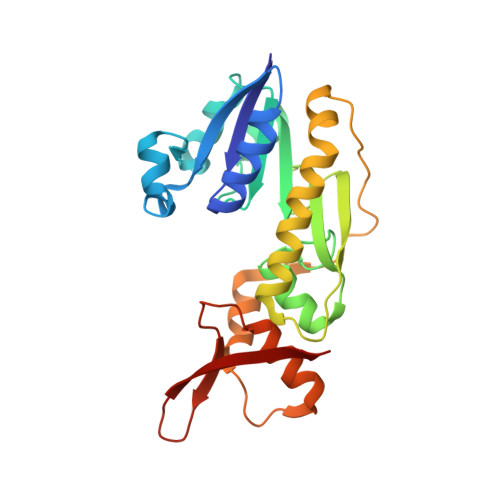
Three-dimensional structure of a sugar N-formyltransferase from Francisella tularensis.

Zimmer, A.L.Thoden, J.B.Holden, H.M.

(2014) Protein Sci 23: 273-283

  • DOI: https://doi.org/10.1002/pro.2409
  • Primary Citation of Related Structures:  
    4NV1

  • PubMed Abstract: 

    N-formylated sugars have been observed on the O-antigens of such pathogenic Gram-negative bacteria as Campylobacter jejuni and Francisella tularensis. Until recently, however, little was known regarding the overall molecular architectures of the N-formyltransferases that are required for the biosynthesis of these unusual sugars. Here we demonstrate that the protein encoded by the wbtj gene from F. tularensis is an N-formyltransferase that functions on dTDP-4-amino-4,6-dideoxy-d-glucose as its substrate. The enzyme, hereafter referred to as WbtJ, demonstrates a strict requirement for N(10) -formyltetrahydrofolate as its carbon source. In addition to the kinetic analysis, the three-dimensional structure of the enzyme was solved in the presence of dTDP-sugar ligands to a nominal resolution of 2.1 Å. Each subunit of the dimeric enzyme is dominated by a "core" domain defined by Met 1 to Ser 185. This core motif harbors the active site residues. Following the core domain, the last 56 residues fold into two α-helices and a β-hairpin motif. The hairpin motif is responsible primarily for the subunit:subunit interface, which is characterized by a rather hydrophobic pocket. From the study presented here, it is now known that WbtJ functions on C-4' amino sugars. Another enzyme recently investigated in the laboratory, WlaRD, formylates only C-3' amino sugars. Strikingly, the quaternary structures of WbtJ and WlaRD are remarkably different. In addition, there are several significant variations in the side chains that line their active site pockets, which may be important for substrate specificity. Details concerning the kinetic and structural properties of WbtJ are presented.


  • Organizational Affiliation

    Department of Biochemistry, University of Wisconsin, Madison, Wisconsin, 53706.


Macromolecules
Find similar proteins by:  (by identity cutoff)  |  3D Structure
Entity ID: 1
MoleculeChains Sequence LengthOrganismDetailsImage
Formyltransferase243Francisella tularensis subsp. tularensis SCHU S4Mutation(s): 0 
Gene Names: wbtJ
UniProt
Find proteins for Q5NF01 (Francisella tularensis subsp. tularensis (strain SCHU S4 / Schu 4))
Explore Q5NF01 
Go to UniProtKB:  Q5NF01
Entity Groups  
Sequence Clusters30% Identity50% Identity70% Identity90% Identity95% Identity100% Identity
UniProt GroupQ5NF01
Sequence Annotations
Expand
  • Reference Sequence
Small Molecules
Ligands 4 Unique
IDChains Name / Formula / InChI Key2D Diagram3D Interactions
4TG
Query on 4TG

Download Ideal Coordinates CCD File 
O [auth B]dTDP-4,6-dideoxy-4-formamido-glucose
C17 H27 N3 O15 P2
QULUVRDLMBXPHO-GJSHGOAISA-N
0FX
Query on 0FX

Download Ideal Coordinates CCD File 
I [auth C],
L [auth E],
Q [auth A],
R [auth G]
dTDP-4-amino-4,6-dideoxyglucose
C16 H27 N3 O14 P2
UIVJXHWSIFBBCY-LPGAPTBISA-N
TYD
Query on TYD

Download Ideal Coordinates CCD File 
K [auth D],
M [auth F],
S [auth H]
THYMIDINE-5'-DIPHOSPHATE
C10 H16 N2 O11 P2
UJLXYODCHAELLY-XLPZGREQSA-N
PO4
Query on PO4

Download Ideal Coordinates CCD File 
J [auth C],
N [auth F],
P [auth B],
T [auth H],
U [auth H]
PHOSPHATE ION
O4 P
NBIIXXVUZAFLBC-UHFFFAOYSA-K
Experimental Data & Validation

Experimental Data

  • Method: X-RAY DIFFRACTION
  • Resolution: 2.10 Å
  • R-Value Free: 0.270 
  • R-Value Work: 0.191 
  • R-Value Observed: 0.195 
  • Space Group: P 1
Unit Cell:
Length ( Å )Angle ( ˚ )
a = 71.942α = 71.72
b = 80.065β = 88.62
c = 109.852γ = 89.78
Software Package:
Software NamePurpose
HKL-3000data collection
SOLVEphasing
REFMACrefinement
HKL-3000data reduction
HKL-3000data scaling

Structure Validation

View Full Validation Report


Ligand Structure Quality Assessment 


Entry History 

Deposition Data

Revision History  (Full details and data files)

  • Version 1.0: 2013-12-25
    Type: Initial release
  • Version 1.1: 2014-03-19
    Changes: Database references
  • Version 1.2: 2024-02-28
    Changes: Data collection, Database references, Derived calculations