4NF9

Structure of the Knl1/Nsl1 complex


Experimental Data Snapshot

  • Method: X-RAY DIFFRACTION
  • Resolution: 2.80 Å
  • R-Value Free: 0.265 
  • R-Value Work: 0.207 
  • R-Value Observed: 0.210 

wwPDB Validation   3D Report Full Report


This is version 1.0 of the entry. See complete history


Literature

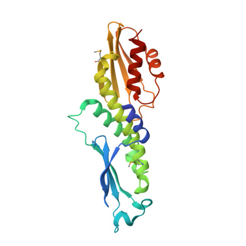
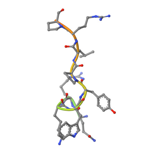
Modular Assembly of RWD Domains on the Mis12 Complex Underlies Outer Kinetochore Organization.

Petrovic, A.Mosalaganti, S.Keller, J.Mattiuzzo, M.Overlack, K.Krenn, V.De Antoni, A.Wohlgemuth, S.Cecatiello, V.Pasqualato, S.Raunser, S.Musacchio, A.

(2014) Mol Cell 53: 591-605

  • DOI: https://doi.org/10.1016/j.molcel.2014.01.019
  • Primary Citation of Related Structures:  
    4NF9, 4NFA

  • PubMed Abstract: 

    Faithful chromosome segregation is mandatory for cell and organismal viability. Kinetochores, large protein assemblies embedded in centromeric chromatin, establish a mechanical link between chromosomes and spindle microtubules. The KMN network, a conserved 10-subunit kinetochore complex, harbors the microtubule-binding interface. RWD domains in the KMN subunits Spc24 and Spc25 mediate kinetochore targeting of the microtubule-binding subunits by interacting with the Mis12 complex, a KMN subcomplex that tethers directly onto the underlying chromatin layer. Here, we show that Knl1, a KMN subunit involved in mitotic checkpoint signaling, also contains RWD domains that bind the Mis12 complex and that mediate kinetochore targeting of Knl1. By reporting the first 3D electron microscopy structure of the KMN network, we provide a comprehensive framework to interpret how interactions of RWD-containing proteins with the Mis12 complex shape KMN network topology. Our observations unveil a regular pattern in the construction of the outer kinetochore.


  • Organizational Affiliation

    Department of Mechanistic Cell Biology, Max Planck Institute of Molecular Physiology, Otto-Hahn-Straße 11, 44227 Dortmund, Germany.


Macromolecules
Find similar proteins by:  (by identity cutoff)  |  3D Structure
Entity ID: 1
MoleculeChains Sequence LengthOrganismDetailsImage
Protein CASC5
A, B
221Homo sapiensMutation(s): 0 
Gene Names: CASC5KIAA1570KNL1
UniProt & NIH Common Fund Data Resources
Find proteins for Q8NG31 (Homo sapiens)
Explore Q8NG31 
Go to UniProtKB:  Q8NG31
PHAROS:  Q8NG31
GTEx:  ENSG00000137812 
Entity Groups  
Sequence Clusters30% Identity50% Identity70% Identity90% Identity95% Identity100% Identity
UniProt GroupQ8NG31
Sequence Annotations
Expand
  • Reference Sequence
Find similar proteins by:  (by identity cutoff)  |  3D Structure
Entity ID: 2
MoleculeChains Sequence LengthOrganismDetailsImage
Kinetochore-associated protein NSL1 homolog
C, D
26Homo sapiensMutation(s): 0 
UniProt & NIH Common Fund Data Resources
Find proteins for Q96IY1 (Homo sapiens)
Explore Q96IY1 
Go to UniProtKB:  Q96IY1
PHAROS:  Q96IY1
GTEx:  ENSG00000117697 
Entity Groups  
Sequence Clusters30% Identity50% Identity70% Identity90% Identity95% Identity100% Identity
UniProt GroupQ96IY1
Sequence Annotations
Expand
  • Reference Sequence
Small Molecules
Ligands 1 Unique
IDChains Name / Formula / InChI Key2D Diagram3D Interactions
CL
Query on CL

Download Ideal Coordinates CCD File 
E [auth A]CHLORIDE ION
Cl
VEXZGXHMUGYJMC-UHFFFAOYSA-M
Modified Residues  1 Unique
IDChains TypeFormula2D DiagramParent
MSE
Query on MSE
A, B
L-PEPTIDE LINKINGC5 H11 N O2 SeMET
Experimental Data & Validation

Experimental Data

  • Method: X-RAY DIFFRACTION
  • Resolution: 2.80 Å
  • R-Value Free: 0.265 
  • R-Value Work: 0.207 
  • R-Value Observed: 0.210 
  • Space Group: P 21 21 21
Unit Cell:
Length ( Å )Angle ( ˚ )
a = 60.71α = 90
b = 71.63β = 90
c = 177.21γ = 90
Software Package:
Software NamePurpose
XDSdata scaling
MOLREPphasing
REFMACrefinement
XDSdata reduction
XSCALEdata scaling

Structure Validation

View Full Validation Report


Entry History 

Deposition Data

Revision History  (Full details and data files)

  • Version 1.0: 2014-03-19
    Type: Initial release