4NDW

Crystal STRUCTURE OF MYCOBACTERIUM TUBERCULOSIS ESX-1 SECRETED PROTEIN REGULATOR (EspR)


Experimental Data Snapshot

  • Method: X-RAY DIFFRACTION
  • Resolution: 3.30 Å
  • R-Value Free: 0.324 
  • R-Value Work: 0.266 
  • R-Value Observed: 0.268 

wwPDB Validation   3D Report Full Report


This is version 1.2 of the entry. See complete history


Literature

Comparison of four different crystal forms of the Mycobacterium tuberculosis ESX-1 secreted protein regulator EspR

Gangwar, S.P.Meena, S.R.Saxena, A.K.

(2014) Acta Crystallogr Sect F Struct Biol Cryst Commun 70: 433-437

  • DOI: https://doi.org/10.1107/S2053230X14004166
  • Primary Citation of Related Structures:  
    4NDW

  • PubMed Abstract: 

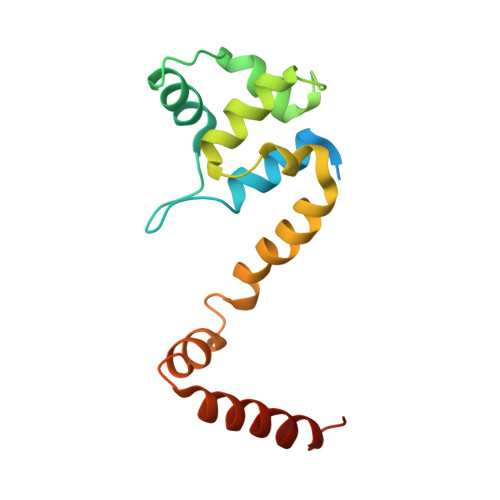
    The Mycobacterium tuberculosis ESX-1 secreted protein regulator (EspR, Rv3849) is the key protein that delivers bacterial proteins into the host cell during mycobacterial infection. EspR binds directly to the espACD operon and is involved in transcriptional activation. In the current study, M. tuberculosis EspR has been crystallized and its X-ray structure has been determined at 3.3 Å resolution in a P3221 crystal form. EspR forms a physiological dimer in the crystal. Each EspR monomer contains an N-terminal helix-turn-helix DNA-binding domain and a C-terminal dimerization domain. The EspR structure in the P3221 crystal form was compared with previously determined EspR structures in P32, P21 and P212121 crystal forms. Structural comparison analysis indicated that the N-terminal helix-turn-helix domain of EspR acquires a rigid structure in the four crystal forms. However, significant structural differences were observed in the C-terminal domain of EspR in the P21 crystal form when compared with the P3221 and P32 crystal forms. The interaction, stabilization energy and buried surface area analysis of EspR in the four different crystal forms have provided information about the physiological dimer interface of EspR.


  • Organizational Affiliation

    School of Life Sciences, Jawaharlal Nehru University, New Delhi 110 067, India.


Macromolecules
Find similar proteins by:  (by identity cutoff)  |  3D Structure
Entity ID: 1
MoleculeChains Sequence LengthOrganismDetailsImage
Nucleoid-associated protein EspR
A, B
153Mycobacterium tuberculosisMutation(s): 0 
Gene Names: espRMT3964Rv3849RV3849C
UniProt
Find proteins for P9WJB7 (Mycobacterium tuberculosis (strain ATCC 25618 / H37Rv))
Explore P9WJB7 
Go to UniProtKB:  P9WJB7
Entity Groups  
Sequence Clusters30% Identity50% Identity70% Identity90% Identity95% Identity100% Identity
UniProt GroupP9WJB7
Sequence Annotations
Expand
  • Reference Sequence
Experimental Data & Validation

Experimental Data

  • Method: X-RAY DIFFRACTION
  • Resolution: 3.30 Å
  • R-Value Free: 0.324 
  • R-Value Work: 0.266 
  • R-Value Observed: 0.268 
  • Space Group: P 32 2 1
Unit Cell:
Length ( Å )Angle ( ˚ )
a = 83.89α = 90
b = 83.89β = 90
c = 130.75γ = 120
Software Package:
Software NamePurpose
MOSFLMdata reduction
PHENIXmodel building
PHENIXrefinement
SCALAdata scaling
PHENIXphasing

Structure Validation

View Full Validation Report


Entry History 

Deposition Data

Revision History  (Full details and data files)

  • Version 1.0: 2014-04-23
    Type: Initial release
  • Version 1.1: 2019-12-18
    Changes: Data collection, Database references
  • Version 1.2: 2024-03-20
    Changes: Data collection, Database references