4NA2

Crystal Structure of the second ketosynthase from the bacillaene polyketide synthase bound to its natural intermediate


Experimental Data Snapshot

  • Method: X-RAY DIFFRACTION
  • Resolution: 2.30 Å
  • R-Value Free: 0.260 
  • R-Value Work: 0.210 
  • R-Value Observed: 0.213 

wwPDB Validation   3D Report Full Report


This is version 1.2 of the entry. See complete history


Literature

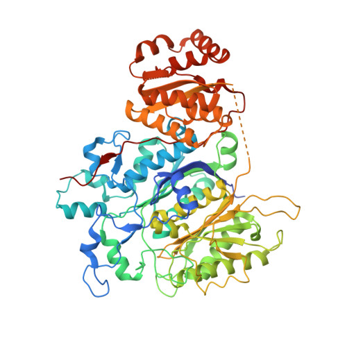
A close look at a ketosynthase from a trans-acyltransferase modular polyketide synthase.

Gay, D.C.Gay, G.Axelrod, A.J.Jenner, M.Kohlhaas, C.Kampa, A.Oldham, N.J.Piel, J.Keatinge-Clay, A.T.

(2014) Structure 22: 444-451

  • DOI: https://doi.org/10.1016/j.str.2013.12.016
  • Primary Citation of Related Structures:  
    4NA1, 4NA2, 4NA3

  • PubMed Abstract: 

    The recently discovered trans-acyltransferase modular polyketide synthases catalyze the biosynthesis of a wide range of bioactive natural products in bacteria. Here we report the structure of the second ketosynthase from the bacillaene trans-acyltransferase polyketide synthase. This 1.95 Å resolution structure provides the highest resolution view available of a modular polyketide synthase ketosynthase and reveals a flanking subdomain that is homologous to an ordered linker in cis-acyltransferase modular polyketide synthases. The structure of the cysteine-to-serine mutant of the ketosynthase acylated by its natural substrate provides high-resolution details of how a native polyketide intermediate is bound and helps explain the basis of ketosynthase substrate specificity. The substrate range of the ketosynthase was further investigated by mass spectrometry.


  • Organizational Affiliation

    Department of Molecular Biosciences, The University of Texas at Austin, 1 University Station A5300, Austin, TX 78712, USA.


Macromolecules
Find similar proteins by:  (by identity cutoff)  |  3D Structure
Entity ID: 1
MoleculeChains Sequence LengthOrganismDetailsImage
Polyketide synthase PksJ
A, B
637Bacillus subtilis subsp. subtilis str. 168Mutation(s): 1 
Gene Names: pksJpksKBSU17180
EC: 2.3.1
UniProt
Find proteins for P40806 (Bacillus subtilis (strain 168))
Explore P40806 
Go to UniProtKB:  P40806
Entity Groups  
Sequence Clusters30% Identity50% Identity70% Identity90% Identity95% Identity100% Identity
UniProt GroupP40806
Sequence Annotations
Expand
  • Reference Sequence
Experimental Data & Validation

Experimental Data

  • Method: X-RAY DIFFRACTION
  • Resolution: 2.30 Å
  • R-Value Free: 0.260 
  • R-Value Work: 0.210 
  • R-Value Observed: 0.213 
  • Space Group: C 1 2 1
Unit Cell:
Length ( Å )Angle ( ˚ )
a = 165.12α = 90
b = 71.34β = 95.1
c = 116.47γ = 90
Software Package:
Software NamePurpose
MAR345dtbdata collection
PHASESphasing
REFMACrefinement
MOSFLMdata reduction
SCALAdata scaling

Structure Validation

View Full Validation Report


Entry History 

Deposition Data

Revision History  (Full details and data files)

  • Version 1.0: 2014-02-19
    Type: Initial release
  • Version 1.1: 2014-03-26
    Changes: Database references
  • Version 1.2: 2023-09-20
    Changes: Data collection, Database references, Derived calculations, Refinement description