4N53

Human enterovirus 71 uncoating intermediate captured at atomic resolution


Experimental Data Snapshot

  • Method: X-RAY DIFFRACTION
  • Resolution: 3.31 Å
  • R-Value Free: 0.261 
  • R-Value Work: 0.228 
  • R-Value Observed: 0.228 

wwPDB Validation   3D Report Full Report


Ligand Structure Quality Assessment 


This is version 1.2 of the entry. See complete history


Literature

Human enterovirus 71 uncoating captured at atomic resolution.

Lyu, K.Ding, J.Han, J.F.Zhang, Y.Wu, X.Y.He, Y.L.Qin, C.F.Chen, R.

(2014) J Virol 88: 3114-3126

  • DOI: https://doi.org/10.1128/JVI.03029-13
  • Primary Citation of Related Structures:  
    4N43, 4N53

  • PubMed Abstract: 

    Human enterovirus 71 (EV71) is the major causative agent of severe hand-foot-and-mouth diseases (HFMD) in young children, and structural characterization of EV71 during its life cycle can aid in the development of therapeutics against HFMD. Here, we present the atomic structures of the full virion and an uncoating intermediate of a clinical EV71 C4 strain to illustrate the structural changes in the full virion that lead to the formation of the uncoating intermediate prepared for RNA release. Although the VP1 N-terminal regions observed to penetrate through the junction channel at the quasi-3-fold axis in the uncoating intermediate of coxsackievirus A16 were not observed in the EV71 uncoating intermediate, drastic conformational changes occur in this region, as has been observed in all capsid proteins. Additionally, the RNA genome interacts with the N-terminal extensions of VP1 and residues 32 to 36 of VP3, both of which are situated at the bottom of the junction. These observations highlight the importance of the junction for genome release. Furthermore, EV71 uncoating is associated with apparent rearrangements and expansion around the 2- and 5-fold axes without obvious changes around the 3-fold axes. Therefore, these structures enabled the identification of hot spots for capsid rearrangements, which led to the hypothesis that the protomer interface near the junction and the 2-fold axis permits the opening of large channels for the exit of polypeptides and viral RNA, which is an uncoating mechanism that is likely conserved in enteroviruses.


  • Organizational Affiliation

    Key Laboratory of Molecular Virology and Immunology, Institut Pasteur of Shanghai, Chinese Academy of Sciences, Shanghai, China.


Macromolecules
Find similar proteins by:  (by identity cutoff)  |  3D Structure
Entity ID: 1
MoleculeChains Sequence LengthOrganismDetailsImage
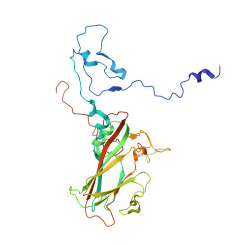
Capsid protein VP1297Enterovirus A71Mutation(s): 0 
UniProt
Find proteins for S5QA87 (Human enterovirus 71)
Explore S5QA87 
Go to UniProtKB:  S5QA87
Entity Groups  
Sequence Clusters30% Identity50% Identity70% Identity90% Identity95% Identity100% Identity
UniProt GroupS5QA87
Sequence Annotations
Expand
  • Reference Sequence
Find similar proteins by:  (by identity cutoff)  |  3D Structure
Entity ID: 2
MoleculeChains Sequence LengthOrganismDetailsImage
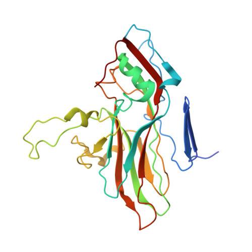
Capsid protein VP2254Enterovirus A71Mutation(s): 0 
UniProt
Find proteins for S4VM80 (Human enterovirus 71)
Explore S4VM80 
Go to UniProtKB:  S4VM80
Entity Groups  
Sequence Clusters30% Identity50% Identity70% Identity90% Identity95% Identity100% Identity
UniProt GroupS4VM80
Sequence Annotations
Expand
  • Reference Sequence
Find similar proteins by:  (by identity cutoff)  |  3D Structure
Entity ID: 3
MoleculeChains Sequence LengthOrganismDetailsImage
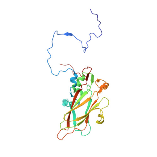
Capsid protein VP3242Enterovirus A71Mutation(s): 0 
UniProt
Find proteins for S5ZCI0 (Human enterovirus 71)
Explore S5ZCI0 
Go to UniProtKB:  S5ZCI0
Entity Groups  
Sequence Clusters30% Identity50% Identity70% Identity90% Identity95% Identity100% Identity
UniProt GroupS5ZCI0
Sequence Annotations
Expand
  • Reference Sequence
Find similar proteins by:  (by identity cutoff)  |  3D Structure
Entity ID: 4
MoleculeChains Sequence LengthOrganismDetailsImage
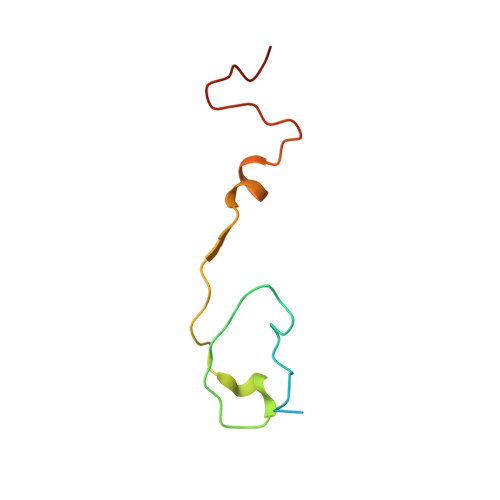
Capsid protein VP469Enterovirus A71Mutation(s): 0 
UniProt
Find proteins for S6C3M9 (Human enterovirus 71)
Explore S6C3M9 
Go to UniProtKB:  S6C3M9
Entity Groups  
Sequence Clusters30% Identity50% Identity70% Identity90% Identity95% Identity100% Identity
UniProt GroupS6C3M9
Sequence Annotations
Expand
  • Reference Sequence
Small Molecules
Ligands 1 Unique
IDChains Name / Formula / InChI Key2D Diagram3D Interactions
SPH
Query on SPH

Download Ideal Coordinates CCD File 
E [auth A]SPHINGOSINE
C18 H37 N O2
WWUZIQQURGPMPG-MSOLQXFVSA-N
Experimental Data & Validation

Experimental Data

  • Method: X-RAY DIFFRACTION
  • Resolution: 3.31 Å
  • R-Value Free: 0.261 
  • R-Value Work: 0.228 
  • R-Value Observed: 0.228 
  • Space Group: I 2 3
Unit Cell:
Length ( Å )Angle ( ˚ )
a = 591.375α = 90
b = 591.375β = 90
c = 591.375γ = 90
Software Package:
Software NamePurpose
SCALEPACKdata scaling
PHENIXrefinement
PDB_EXTRACTdata extraction
HKL-2000data collection
HKL-2000data reduction
PHENIXphasing

Structure Validation

View Full Validation Report


Ligand Structure Quality Assessment 


Entry History 

Deposition Data

Revision History  (Full details and data files)

  • Version 1.0: 2014-02-05
    Type: Initial release
  • Version 1.1: 2014-03-12
    Changes: Database references
  • Version 1.2: 2023-09-20
    Changes: Data collection, Database references, Derived calculations, Refinement description