4N4O

Nitrosomonas europea HAO soaked in NH2OH


Experimental Data Snapshot

  • Method: X-RAY DIFFRACTION
  • Resolution: 2.47 Å
  • R-Value Free: 0.273 
  • R-Value Work: 0.224 
  • R-Value Observed: 0.229 

wwPDB Validation   3D Report Full Report


Ligand Structure Quality Assessment 


This is version 2.1 of the entry. See complete history


Literature

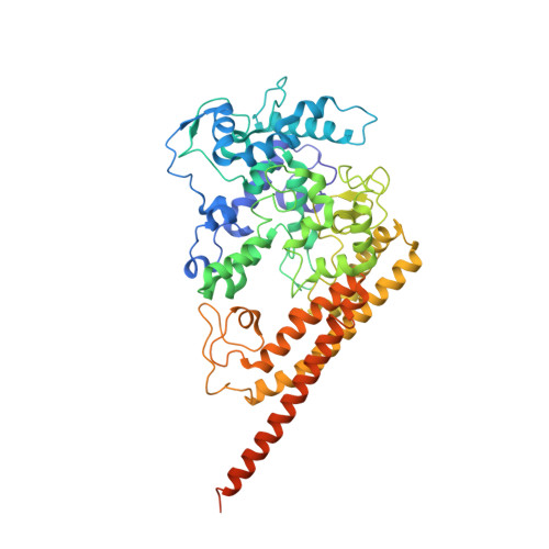
Structural Basis of Biological NO Generation by Octaheme Oxidoreductases.

Maalcke, W.J.Dietl, A.Marritt, S.J.Butt, J.N.Jetten, M.S.Keltjens, J.T.Barends, T.R.Kartal, B.

(2014) J Biol Chem 289: 1228-1242

  • DOI: https://doi.org/10.1074/jbc.M113.525147
  • Primary Citation of Related Structures:  
    4N4J, 4N4K, 4N4L, 4N4M, 4N4N, 4N4O

  • PubMed Abstract: 

    Nitric oxide is an important molecule in all domains of life with significant biological functions in both pro- and eukaryotes. Anaerobic ammonium-oxidizing (anammox) bacteria that contribute substantially to the release of fixed nitrogen into the atmosphere use the oxidizing power of NO to activate inert ammonium into hydrazine (N2H4). Here, we describe an enzyme from the anammox bacterium Kuenenia stuttgartiensis that uses a novel pathway to make NO from hydroxylamine. This new enzyme is related to octaheme hydroxylamine oxidoreductase, a key protein in aerobic ammonium-oxidizing bacteria. By a multiphasic approach including the determination of the crystal structure of the K. stuttgartiensis enzyme at 1.8 Å resolution and refinement and reassessment of the hydroxylamine oxidoreductase structure from Nitrosomonas europaea, both in the presence and absence of their substrates, we propose a model for NO formation by the K. stuttgartiensis enzyme. Our results expand the understanding of the functions that the widespread family of octaheme proteins have.


  • Organizational Affiliation

    From the Department of Microbiology, Institute for Water and Wetland Research, Radboud University Nijmegen, 6525AJ Nijmegen, The Netherlands.


Macromolecules
Find similar proteins by:  (by identity cutoff)  |  3D Structure
Entity ID: 1
MoleculeChains Sequence LengthOrganismDetailsImage
Hydroxylamine oxidoreductase
A, C, E
546Nitrosomonas europaeaMutation(s): 0 
EC: 1.7.2.6
UniProt
Find proteins for Q50925 (Nitrosomonas europaea (strain ATCC 19718 / CIP 103999 / KCTC 2705 / NBRC 14298))
Explore Q50925 
Go to UniProtKB:  Q50925
Entity Groups  
Sequence Clusters30% Identity50% Identity70% Identity90% Identity95% Identity100% Identity
UniProt GroupQ50925
Sequence Annotations
Expand
  • Reference Sequence
Find similar proteins by:  (by identity cutoff)  |  3D Structure
Entity ID: 2
MoleculeChains Sequence LengthOrganismDetailsImage
hydroxylamine oxidoreductase
B, D, F
57Nitrosomonas europaeaMutation(s): 0 
EC: 1.7.2.6
UniProt
Find proteins for Q82V11 (Nitrosomonas europaea (strain ATCC 19718 / CIP 103999 / KCTC 2705 / NBRC 14298))
Explore Q82V11 
Go to UniProtKB:  Q82V11
Entity Groups  
Sequence Clusters30% Identity50% Identity70% Identity90% Identity95% Identity100% Identity
UniProt GroupQ82V11
Sequence Annotations
Expand
  • Reference Sequence
Small Molecules
Ligands 5 Unique
IDChains Name / Formula / InChI Key2D Diagram3D Interactions
HEC
Query on HEC

Download Ideal Coordinates CCD File 
BA [auth E]
CA [auth E]
DA [auth E]
EA [auth E]
FA [auth E]
BA [auth E],
CA [auth E],
DA [auth E],
EA [auth E],
FA [auth E],
G [auth A],
GA [auth E],
H [auth A],
HA [auth E],
I [auth A],
IA [auth E],
J [auth A],
K [auth A],
L [auth A],
M [auth A],
N [auth A],
R [auth C],
S [auth C],
T [auth C],
U [auth C],
V [auth C],
W [auth C],
X [auth C],
Y [auth C]
HEME C
C34 H34 Fe N4 O4
HXQIYSLZKNYNMH-LJNAALQVSA-N
12P
Query on 12P

Download Ideal Coordinates CCD File 
AA [auth C],
Q [auth A]
DODECAETHYLENE GLYCOL
C24 H50 O13
WRZXKWFJEFFURH-UHFFFAOYSA-N
PO4
Query on PO4

Download Ideal Coordinates CCD File 
JA [auth E]PHOSPHATE ION
O4 P
NBIIXXVUZAFLBC-UHFFFAOYSA-K
K
Query on K

Download Ideal Coordinates CCD File 
O [auth A]POTASSIUM ION
K
NPYPAHLBTDXSSS-UHFFFAOYSA-N
HOA
Query on HOA

Download Ideal Coordinates CCD File 
KA [auth E],
P [auth A],
Z [auth C]
HYDROXYAMINE
H3 N O
AVXURJPOCDRRFD-UHFFFAOYSA-N
Experimental Data & Validation

Experimental Data

  • Method: X-RAY DIFFRACTION
  • Resolution: 2.47 Å
  • R-Value Free: 0.273 
  • R-Value Work: 0.224 
  • R-Value Observed: 0.229 
  • Space Group: P 21 21 2
Unit Cell:
Length ( Å )Angle ( ˚ )
a = 140.09α = 90
b = 141.96β = 90
c = 106.48γ = 90
Software Package:
Software NamePurpose
HEIDIdata collection
REFMACrefinement
XDSdata reduction
XSCALEdata scaling
REFMACphasing

Structure Validation

View Full Validation Report


Ligand Structure Quality Assessment 


Entry History 

Deposition Data

Revision History  (Full details and data files)

  • Version 1.0: 2013-12-11
    Type: Initial release
  • Version 1.1: 2014-01-15
    Changes: Database references
  • Version 1.2: 2014-02-05
    Changes: Database references
  • Version 2.0: 2021-03-10
    Changes: Atomic model, Data collection, Derived calculations, Non-polymer description, Structure summary
  • Version 2.1: 2023-09-20
    Changes: Data collection, Database references, Refinement description