4MZB

Crystal structure of Grx1 from Plasmodium falciparum


Experimental Data Snapshot

  • Method: X-RAY DIFFRACTION
  • Resolution: 1.04 Å
  • R-Value Free: 0.129 
  • R-Value Work: 0.117 
  • R-Value Observed: 0.118 

wwPDB Validation   3D Report Full Report


Ligand Structure Quality Assessment 


This is version 1.4 of the entry. See complete history


Literature

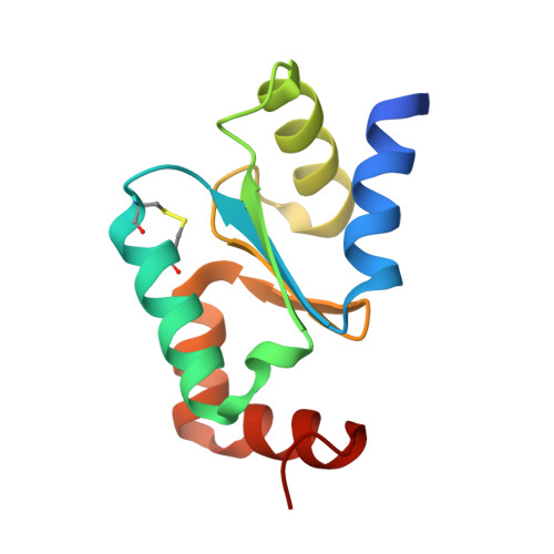
Atomic resolution crystal structure of glutaredoxin 1 from Plasmodium falciparum and comparison with other glutaredoxins.

Yogavel, M.Tripathi, T.Gupta, A.Banday, M.M.Rahlfs, S.Becker, K.Belrhali, H.Sharma, A.

(2014) Acta Crystallogr D Biol Crystallogr 70: 91-100

  • DOI: https://doi.org/10.1107/S1399004713025285
  • Primary Citation of Related Structures:  
    4HJM, 4MZB, 4MZC

  • PubMed Abstract: 

    Glutaredoxins (Grxs) are redox proteins that use glutathione ((γ)Glu-Cys-Gly; GSH) as a cofactor. Plasmodium falciparum has one classic dithiol (CXXC) glutaredoxin (glutaredoxin 1; PfGrx1) and three monothiol (CXXS) Grx-like proteins (GLPs), which have five residue insertions prior to the active-site Cys. Here, the crystal structure of PfGrx1 has been determined by the sulfur single-wavelength anomalous diffraction (S-SAD) method utilizing intrinsic protein and solvent S atoms. Several residues were modelled with alternate conformations, and an alternate position was refined for the active-site Cys29 owing to radiation damage. The GSH-binding site is occupied by water polygons and buffer molecules. Structural comparison of PfGrx1 with other Grxs and Grx-like proteins revealed that the GSH-binding motifs (CXXC/CXXS, TVP, CDD, Lys26 and Gln/Arg63) are structurally conserved. Both the monothiol and dithiol Grxs possess three conserved water molecules; two of these were located in the GSH-binding site. PfGrx1 has several polar and charged amino-acid substitutions that provide structurally important additional hydrogen bonds and salt bridges missing in other Grxs.


  • Organizational Affiliation

    Structural and Computational Biology Group, International Centre for Genetic Engineering and Biotechnology (ICGEB), Aruna Asaf Ali Road, New Delhi 110 067, India.


Macromolecules
Find similar proteins by:  (by identity cutoff)  |  3D Structure
Entity ID: 1
MoleculeChains Sequence LengthOrganismDetailsImage
Glutaredoxin111Plasmodium falciparum 3D7Mutation(s): 0 
Gene Names: GRX1PFC0271c
EC: 1.20.4.1
UniProt
Find proteins for Q9NLB2 (Plasmodium falciparum (isolate 3D7))
Explore Q9NLB2 
Go to UniProtKB:  Q9NLB2
Entity Groups  
Sequence Clusters30% Identity50% Identity70% Identity90% Identity95% Identity100% Identity
UniProt GroupQ9NLB2
Sequence Annotations
Expand
  • Reference Sequence
Small Molecules
Ligands 2 Unique
IDChains Name / Formula / InChI Key2D Diagram3D Interactions
MPO
Query on MPO

Download Ideal Coordinates CCD File 
B [auth A]3[N-MORPHOLINO]PROPANE SULFONIC ACID
C7 H15 N O4 S
DVLFYONBTKHTER-UHFFFAOYSA-N
MPD
Query on MPD

Download Ideal Coordinates CCD File 
C [auth A](4S)-2-METHYL-2,4-PENTANEDIOL
C6 H14 O2
SVTBMSDMJJWYQN-YFKPBYRVSA-N
Experimental Data & Validation

Experimental Data

  • Method: X-RAY DIFFRACTION
  • Resolution: 1.04 Å
  • R-Value Free: 0.129 
  • R-Value Work: 0.117 
  • R-Value Observed: 0.118 
  • Space Group: P 32 2 1
Unit Cell:
Length ( Å )Angle ( ˚ )
a = 47.953α = 90
b = 47.953β = 90
c = 82.45γ = 120
Software Package:
Software NamePurpose
MOLREPphasing
PHENIXrefinement
PDB_EXTRACTdata extraction
DNAdata collection
HKL-2000data reduction
HKL-2000data scaling

Structure Validation

View Full Validation Report


Ligand Structure Quality Assessment 


Entry History 

Deposition Data

Revision History  (Full details and data files)

  • Version 1.0: 2013-10-09
    Type: Initial release
  • Version 1.1: 2014-01-08
    Changes: Database references
  • Version 1.2: 2014-09-24
    Changes: Database references
  • Version 1.3: 2020-09-09
    Changes: Derived calculations, Structure summary
  • Version 1.4: 2023-09-20
    Changes: Data collection, Database references, Refinement description