


4MOI
Pyranose 2-oxidase H450G/V546C double mutant with 3-fluorinated glucose
- PDB DOI: https://doi.org/10.2210/pdb4MOI/pdb
- Classification: OXIDOREDUCTASE
- Organism(s): Trametes ochracea
- Expression System: Escherichia coli BL21(DE3)
- Mutation(s): Yes
- Deposited: 2013-09-12 Released: 2014-02-05
Experimental Data Snapshot
- Method: X-RAY DIFFRACTION
- Resolution: 1.90 Å
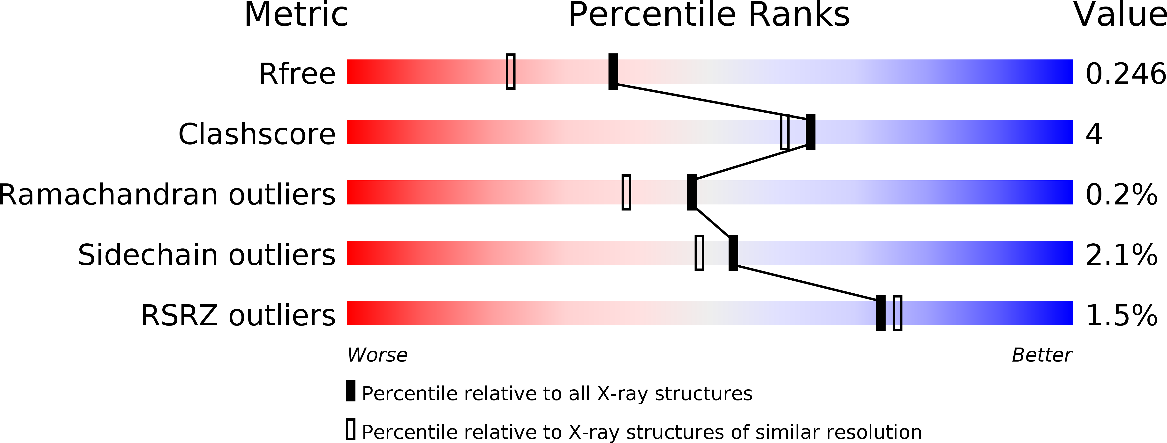
- R-Value Free: 0.245
- R-Value Work: 0.196
- R-Value Observed: 0.197
This is version 1.2 of the entry. See complete history.

Macromolecules
Find similar proteins by:
(by identity cutoff) | 3D Structure
Entity ID: 1 | |||||
|---|---|---|---|---|---|
| Molecule | Chains | Sequence Length | Organism | Details | Image |
| Pyranose 2-oxidase | 633 | Trametes ochracea | Mutation(s): 2 Gene Names: p2o EC: 1.1.3.10 |  | |
UniProt | |||||
Find proteins for Q7ZA32 (Trametes ochracea) Explore Q7ZA32 Go to UniProtKB: Q7ZA32 | |||||
Entity Groups | |||||
| Sequence Clusters | 30% Identity50% Identity70% Identity90% Identity95% Identity100% Identity | ||||
| UniProt Group | Q7ZA32 | ||||
Sequence AnnotationsExpand | |||||
| |||||
Small Molecules
| Ligands 2 Unique | |||||
|---|---|---|---|---|---|
| ID | Chains | Name / Formula / InChI Key | 2D Diagram | 3D Interactions | |
| FDA Query on FDA | C [auth A], E [auth B] | DIHYDROFLAVINE-ADENINE DINUCLEOTIDE C27 H35 N9 O15 P2 YPZRHBJKEMOYQH-UYBVJOGSSA-N |  | ||
| G3F Query on G3F | D [auth A], F [auth B] | 3-deoxy-3-fluoro-beta-D-glucopyranose C6 H11 F O5 BUMRBAMACDBPKO-AIECOIEWSA-N |  | ||
Experimental Data & Validation
Experimental Data
- Method: X-RAY DIFFRACTION
- Resolution: 1.90 Å
- R-Value Free: 0.245
- R-Value Work: 0.196
- R-Value Observed: 0.197
- Space Group: P 41 21 2
Unit Cell:
| Length ( Å ) | Angle ( ˚ ) |
|---|---|
| a = 101.504 | α = 90 |
| b = 101.504 | β = 90 |
| c = 238.672 | γ = 90 |
| Software Name | Purpose |
|---|---|
| MxCuBE | data collection |
| PHASER | phasing |
| REFMAC | refinement |
| XDS | data reduction |
| XSCALE | data scaling |
Entry History
Deposition Data
- Released Date: 2014-02-05 Deposition Author(s): Tan, T.C., Spadiut, O., Gandini, R., Haltrich, D., Divne, C.
Revision History (Full details and data files)
- Version 1.0: 2014-02-05
Type: Initial release - Version 1.1: 2018-03-07
Changes: Data collection - Version 1.2: 2020-07-29
Type: Remediation
Reason: Carbohydrate remediation
Changes: Data collection, Database references, Derived calculations
