4MO2

Crystal Structure of UDP-N-acetylgalactopyranose mutase from Campylobacter jejuni


Experimental Data Snapshot

  • Method: X-RAY DIFFRACTION
  • Resolution: 2.00 Å
  • R-Value Free: 0.212 
  • R-Value Work: 0.171 
  • R-Value Observed: 0.173 

wwPDB Validation   3D Report Full Report


Ligand Structure Quality Assessment 


This is version 1.2 of the entry. See complete history


Literature

Specificity of a UDP-GalNAc Pyranose-Furanose Mutase: A Potential Therapeutic Target for Campylobacter jejuni Infections.

Poulin, M.B.Shi, Y.Protsko, C.Dalrymple, S.A.Sanders, D.A.Pinto, B.M.Lowary, T.L.

(2014) Chembiochem 15: 47-56

  • DOI: https://doi.org/10.1002/cbic.201300653
  • Primary Citation of Related Structures:  
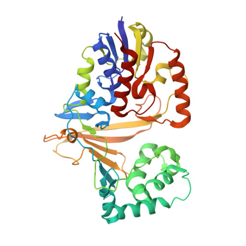
    4MO2

  • PubMed Abstract: 

    Pyranose-furanose mutases are essential enzymes in the life cycle of a number of microorganisms, but are absent in mammalian systems, and hence represent novel targets for drug development. To date, all such mutases show preferential recognition of a single substrate (e.g., UDP-Gal). We report here the detailed structural characterization of the first bifunctional pyranose-furanose mutase, which recognizes both UDP-Gal and UDP-GalNAc. The enzyme under investigation (cjUNGM) is involved in the biosynthesis of capsular polysaccharides (CPSs) in Campylobacter jejuni 11168. These CPSs are known virulence factors that are required for adhesion and invasion of human epithelial cells. Using a combination of UV/visible spectroscopy, X-ray crystallography, saturation transfer difference NMR spectroscopy, molecular dynamics and CORCEMA-ST calculations, we have characterized the binding of the enzyme to both UDP-Galp and UDP-GalpNAc, and compared these interactions with those of a homologous monofunctional mutase enzyme from E. coli (ecUGM). These studies reveal that two arginines in cjUNGM, Arg59 and Arg168, play critical roles in the catalytic mechanism of the enzyme and in controlling its specificity to ultimately lead to a GalfNAc-containing CPS. In ecUGM, these arginines are replaced with histidine and lysine, respectively, and this results in an enzyme that is selective for UDP-Gal. We propose that these changes in amino acids allow C. jejuni 11168 to produce suitable quantities of the sugar nucleotide substrate required for the assembly of a CPS containing GalfNAc, which is essential for viability.


  • Organizational Affiliation

    Alberta Glycomics Centre and Department of Chemistry, The University of Alberta, Gunning-Lemieux Chemistry Centre, 11227 Saskatchewan Drive, Edmonton, AB T6G 2G2 (Canada).


Macromolecules
Find similar proteins by:  (by identity cutoff)  |  3D Structure
Entity ID: 1
MoleculeChains Sequence LengthOrganismDetailsImage
UDP-galactopyranose mutaseA [auth B],
B [auth A]
368Campylobacter jejuni subsp. jejuni NCTC 11168 = ATCC 700819Mutation(s): 0 
Gene Names: Cj1439cglf
EC: 5.4.99.9
UniProt
Find proteins for Q0P8H5 (Campylobacter jejuni subsp. jejuni serotype O:2 (strain ATCC 700819 / NCTC 11168))
Explore Q0P8H5 
Go to UniProtKB:  Q0P8H5
Entity Groups  
Sequence Clusters30% Identity50% Identity70% Identity90% Identity95% Identity100% Identity
UniProt GroupQ0P8H5
Sequence Annotations
Expand
  • Reference Sequence
Small Molecules
Ligands 5 Unique
IDChains Name / Formula / InChI Key2D Diagram3D Interactions
FDA
Query on FDA

Download Ideal Coordinates CCD File 
D [auth B]DIHYDROFLAVINE-ADENINE DINUCLEOTIDE
C27 H35 N9 O15 P2
YPZRHBJKEMOYQH-UYBVJOGSSA-N
FAD
Query on FAD

Download Ideal Coordinates CCD File 
J [auth A]FLAVIN-ADENINE DINUCLEOTIDE
C27 H33 N9 O15 P2
VWWQXMAJTJZDQX-UYBVJOGSSA-N
SO4
Query on SO4

Download Ideal Coordinates CCD File 
C [auth B],
G [auth A]
SULFATE ION
O4 S
QAOWNCQODCNURD-UHFFFAOYSA-L
GOL
Query on GOL

Download Ideal Coordinates CCD File 
E [auth B],
F [auth A]
GLYCEROL
C3 H8 O3
PEDCQBHIVMGVHV-UHFFFAOYSA-N
NH4
Query on NH4

Download Ideal Coordinates CCD File 
H [auth A],
I [auth A]
AMMONIUM ION
H4 N
QGZKDVFQNNGYKY-UHFFFAOYSA-O
Experimental Data & Validation

Experimental Data

  • Method: X-RAY DIFFRACTION
  • Resolution: 2.00 Å
  • R-Value Free: 0.212 
  • R-Value Work: 0.171 
  • R-Value Observed: 0.173 
  • Space Group: P 21 21 21
Unit Cell:
Length ( Å )Angle ( ˚ )
a = 48.42α = 90
b = 116.15β = 90
c = 165.24γ = 90
Software Package:
Software NamePurpose
XSCALEdata scaling
MOLREPphasing
PHENIXrefinement
PDB_EXTRACTdata extraction
MxDCdata collection

Structure Validation

View Full Validation Report


Ligand Structure Quality Assessment 


Entry History 

Deposition Data

Revision History  (Full details and data files)

  • Version 1.0: 2014-01-01
    Type: Initial release
  • Version 1.1: 2017-11-15
    Changes: Refinement description
  • Version 1.2: 2024-02-28
    Changes: Data collection, Database references, Derived calculations