4MLL

The 1.4 A structure of the class D beta-lactamase OXA-1 K70D complexed with oxacillin


Experimental Data Snapshot

  • Method: X-RAY DIFFRACTION
  • Resolution: 1.37 Å
  • R-Value Free: 0.196 
  • R-Value Work: 0.163 
  • R-Value Observed: 0.165 

wwPDB Validation   3D Report Full Report


Ligand Structure Quality Assessment 


This is version 1.2 of the entry. See complete history


Literature

Structural Origins of Oxacillinase Specificity in Class D beta-Lactamases.

June, C.M.Vallier, B.C.Bonomo, R.A.Leonard, D.A.Powers, R.A.

(2014) Antimicrob Agents Chemother 58: 333-341

  • DOI: https://doi.org/10.1128/AAC.01483-13
  • Primary Citation of Related Structures:  
    4MLL

  • PubMed Abstract: 

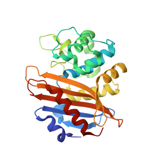
    Since the discovery and use of penicillin, the increase of antibiotic resistance among bacterial pathogens has become a major health concern. The most prevalent resistance mechanism in Gram-negative bacteria is due to β-lactamase expression. Class D β-lactamases are of particular importance due to their presence in multidrug-resistant Acinetobacter baumannii and Pseudomonas aeruginosa. The class D enzymes were initially characterized by their ability to efficiently hydrolyze isoxazolyl-type β-lactams like oxacillin. Due to this substrate preference, these enzymes are traditionally referred to as oxacillinases or OXAs. However, this class is comprised of subfamilies characterized by diverse activities that include oxacillinase, carbapenemase, or cephalosporinase substrate specificity. OXA-1 represents one subtype of class D enzyme that efficiently hydrolyzes oxacillin, and OXA-24/40 represents another with weak oxacillinase, but increased carbapenemase, activity. To examine the structural basis for the substrate selectivity differences between OXA-1 and OXA-24/40, the X-ray crystal structures of deacylation-deficient mutants of these enzymes (Lys70Asp for OXA-1; Lys84Asp for OXA-24) in complexes with oxacillin were determined to 1.4 Å and 2.4 Å, respectively. In the OXA-24/40/oxacillin structure, the hydrophobic R1 side chain of oxacillin disrupts the bridge between Tyr112 and Met223 present in the apo OXA-24/40 structure, causing the main chain of the Met223-containing loop to adopt a completely different conformation. In contrast, in the OXA-1/oxacillin structure, a hydrophobic pocket consisting of Trp102, Met99, Phe217, Leu161, and Leu255 nicely complements oxacillin's nonpolar R1 side chain. Comparison of the OXA-1/oxacillin and OXA-24/40/oxacillin complexes provides novel insight on how substrate selectivity is achieved among subtypes of class D β-lactamases. By elucidating important active site interactions, these findings can also inform the design of novel antibiotics and inhibitors.


  • Organizational Affiliation

    Department of Chemistry, Grand Valley State University, Allendale, Michigan, USA.


Macromolecules
Find similar proteins by:  (by identity cutoff)  |  3D Structure
Entity ID: 1
MoleculeChains Sequence LengthOrganismDetailsImage
Beta-lactamase OXA-1
A, B, C, D
251Escherichia coliMutation(s): 1 
Gene Names: blabla oxa1oxa1
EC: 3.5.2.6
UniProt
Find proteins for P13661 (Escherichia coli)
Explore P13661 
Go to UniProtKB:  P13661
Entity Groups  
Sequence Clusters30% Identity50% Identity70% Identity90% Identity95% Identity100% Identity
UniProt GroupP13661
Sequence Annotations
Expand
  • Reference Sequence
Small Molecules
Ligands 3 Unique
IDChains Name / Formula / InChI Key2D Diagram3D Interactions
1S6
Query on 1S6

Download Ideal Coordinates CCD File 
E [auth A],
J [auth B],
L [auth C],
O [auth D]
(2R,4S)-5,5-dimethyl-2-[(1R)-1-{[(5-methyl-3-phenyl-1,2-oxazol-4-yl)carbonyl]amino}-2-oxoethyl]-1,3-thiazolidine-4-carb oxylic acid
C19 H21 N3 O5 S
QXXMSXCGXIRLPM-ISTRZQFTSA-N
MPD
Query on MPD

Download Ideal Coordinates CCD File 
I [auth A],
Q [auth D],
R [auth D]
(4S)-2-METHYL-2,4-PENTANEDIOL
C6 H14 O2
SVTBMSDMJJWYQN-YFKPBYRVSA-N
PO4
Query on PO4

Download Ideal Coordinates CCD File 
F [auth A]
G [auth A]
H [auth A]
K [auth B]
M [auth C]
F [auth A],
G [auth A],
H [auth A],
K [auth B],
M [auth C],
N [auth C],
P [auth D]
PHOSPHATE ION
O4 P
NBIIXXVUZAFLBC-UHFFFAOYSA-K
Experimental Data & Validation

Experimental Data

  • Method: X-RAY DIFFRACTION
  • Resolution: 1.37 Å
  • R-Value Free: 0.196 
  • R-Value Work: 0.163 
  • R-Value Observed: 0.165 
  • Space Group: P 1
Unit Cell:
Length ( Å )Angle ( ˚ )
a = 50.919α = 80.9
b = 72.609β = 69.87
c = 73.446γ = 71.44
Software Package:
Software NamePurpose
HKL-2000data collection
PHASERphasing
REFMACrefinement
HKL-2000data reduction
HKL-2000data scaling

Structure Validation

View Full Validation Report


Ligand Structure Quality Assessment 


Entry History 

Deposition Data

Revision History  (Full details and data files)

  • Version 1.0: 2013-10-09
    Type: Initial release
  • Version 1.1: 2014-01-08
    Changes: Database references
  • Version 1.2: 2023-09-20
    Changes: Data collection, Database references, Derived calculations, Refinement description, Structure summary