4MH3

Crystal structure of Bovine Mitochondrial Peroxiredoxin III


Experimental Data Snapshot

  • Method: X-RAY DIFFRACTION
  • Resolution: 2.40 Å
  • R-Value Free: 0.221 
  • R-Value Work: 0.182 
  • R-Value Observed: 0.184 

wwPDB Validation   3D Report Full Report


This is version 1.3 of the entry. See complete history


Literature

Improved Catenated Structures of Bovine Peroxiredoxin III F190L Reveal Details of Ring-Ring Interactions and a Novel Conformational State.

Cao, Z.McGow, D.P.Shepherd, C.Lindsay, J.G.

(2015) PLoS One 10: e0123303-e0123303

  • DOI: https://doi.org/10.1371/journal.pone.0123303
  • Primary Citation of Related Structures:  
    4MH2, 4MH3

  • PubMed Abstract: 

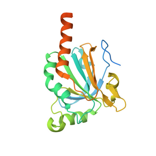
    Mitochondrial 2-cys peroxiredoxin III (PrxIII) is a key player in antioxidant defence reducing locally-generated H2O2 to H2O. A Phe to Leu (F190L) mutation in the C-terminal α-helix of PrxIII, mimicking that found in some bacteria and parasites, increases its resistance to hyperoxidation but has no obvious influence on peroxidase activity. Here we report on the oxidized and reduced crystal structures of bovine PrxIII F190L at 2.4 Å and 2.2 Å, respectively. Both structures exist as two-ring catenanes with their dodecameric rings inclined at 55o to each other, similar to that previously reported for PrxIII C168S. The new higher-resolution structures reveal details of the complex network of H-bonds stabilising the inter-toroid contacts. In addition, Arg123, the key conserved residue, that normally interacts with the catalytic cys (Cp, cys 47) is found in a distinct conformation extending away from the Cp while the characteristic Arg-Glu-Arg network, underpinning the active-site geometry also displays a distinctive arrangement, not observed previously. This novel active-site organisation may provide new insights into the dynamics of the large-scale conformational changes occurring between oxidized and reduced states.


  • Organizational Affiliation

    From the Institute of Molecular, Cell and Systems Biology, CMVLS, University of Glasgow, Glasgow, United Kingdom.


Macromolecules
Find similar proteins by:  (by identity cutoff)  |  3D Structure
Entity ID: 1
MoleculeChains Sequence LengthOrganismDetailsImage
Thioredoxin-dependent peroxide reductase, mitochondrial
A, B, C, D, E
A, B, C, D, E, F, G, H, I, J, K, L
220Bos taurusMutation(s): 1 
Gene Names: AOP1PRDX3
EC: 1.11.1.15
UniProt
Find proteins for P35705 (Bos taurus)
Explore P35705 
Go to UniProtKB:  P35705
Entity Groups  
Sequence Clusters30% Identity50% Identity70% Identity90% Identity95% Identity100% Identity
UniProt GroupP35705
Sequence Annotations
Expand
  • Reference Sequence
Experimental Data & Validation

Experimental Data

  • Method: X-RAY DIFFRACTION
  • Resolution: 2.40 Å
  • R-Value Free: 0.221 
  • R-Value Work: 0.182 
  • R-Value Observed: 0.184 
  • Space Group: P 21 21 2
Unit Cell:
Length ( Å )Angle ( ˚ )
a = 142.895α = 90
b = 290.885β = 90
c = 81.139γ = 90
Software Package:
Software NamePurpose
DNAdata collection
PHASERphasing
REFMACrefinement
MOSFLMdata reduction
SCALAdata scaling

Structure Validation

View Full Validation Report


Entry History 

Deposition Data

Revision History  (Full details and data files)

  • Version 1.0: 2015-03-04
    Type: Initial release
  • Version 1.1: 2015-05-06
    Changes: Database references
  • Version 1.2: 2020-09-09
    Changes: Database references, Derived calculations, Structure summary
  • Version 1.3: 2023-09-20
    Changes: Data collection, Database references, Refinement description