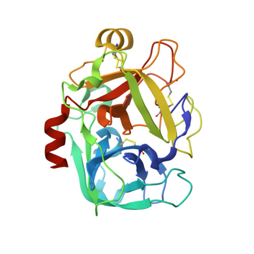
Conformational flexibility in the catalytic triad revealed by the high-resolution crystal structure of Streptomyces erythraeus trypsin in an unliganded state.
Blankenship, E., Vukoti, K., Miyagi, M., Lodowski, D.T.(2014) Acta Crystallogr D Biol Crystallogr 70: 833-840
- PubMed: 24598752
- DOI: https://doi.org/10.1107/S1399004713033658
- Primary Citation of Related Structures:
4M7G - PubMed Abstract:
With more than 500 crystal structures determined, serine proteases make up greater than one-third of all proteases structurally examined to date, making them among the best biochemically and structurally characterized enzymes. Despite the numerous crystallographic and biochemical studies of trypsin and related serine proteases, there are still considerable shortcomings in the understanding of their catalytic mechanism. Streptomyces erythraeus trypsin (SET) does not exhibit autolysis and crystallizes readily at physiological pH; hence, it is well suited for structural studies aimed at extending the understanding of the catalytic mechanism of serine proteases. While X-ray crystallographic structures of this enzyme have been reported, no coordinates have ever been made available in the Protein Data Bank. Based on this, and observations on the extreme stability and unique properties of this particular trypsin, it was decided to crystallize it and determine its structure. Here, the first sub-angstrom resolution structure of an unmodified, unliganded trypsin crystallized at physiological pH is reported. Detailed structural analysis reveals the geometry and structural rigidity of the catalytic triad in the unoccupied active site and comparison to related serine proteases provides a context for interpretation of biochemical studies of catalytic mechanism and activity.
Organizational Affiliation:
Case Center for Proteomics and Bioinformatics, Case Western Reserve University, 10900 Euclid Avenue, Cleveland, OH 44106, USA.