


Structure of a GH39 Beta-xylosidase from Caulobacter crescentus
Polo, C.C., Santos, C.R., Correa, J.M., Simao, R.C.G., Seixas, F.A.V., Murakami, M.T.(null) Thesis
Experimental Data Snapshot
wwPDB Validation 3D Report Full Report

(null) Thesis
Entity ID: 1 | |||||
|---|---|---|---|---|---|
| Molecule | Chains | Sequence Length | Organism | Details | Image |
| Beta-xylosidase | 500 | Caulobacter vibrioides CB15 | Mutation(s): 0 Gene Names: Itgav, CC_2357 | 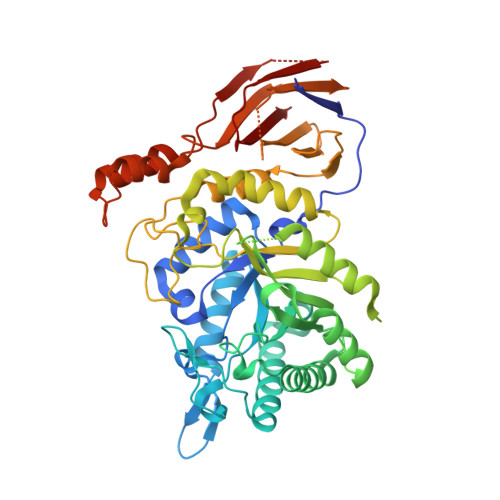 | |
UniProt | |||||
Find proteins for Q9A5U0 (Caulobacter vibrioides (strain ATCC 19089 / CB15)) Explore Q9A5U0 Go to UniProtKB: Q9A5U0 | |||||
Entity Groups | |||||
| Sequence Clusters | 30% Identity50% Identity70% Identity90% Identity95% Identity100% Identity | ||||
| UniProt Group | Q9A5U0 | ||||
Sequence AnnotationsExpand | |||||
| |||||
| Ligands 1 Unique | |||||
|---|---|---|---|---|---|
| ID | Chains | Name / Formula / InChI Key | 2D Diagram | 3D Interactions | |
| MES Query on MES | B [auth A] | 2-(N-MORPHOLINO)-ETHANESULFONIC ACID C6 H13 N O4 S SXGZJKUKBWWHRA-UHFFFAOYSA-N |  | ||
| Length ( Å ) | Angle ( ˚ ) |
|---|---|
| a = 42.287 | α = 90 |
| b = 57.347 | β = 91.38 |
| c = 94.05 | γ = 90 |
| Software Name | Purpose |
|---|---|
| NatXray | data collection |
| PHASER | phasing |
| REFMAC | refinement |
| DENZO | data reduction |
| SCALEPACK | data scaling |
RCSB PDB (citation) is hosted by
RCSB PDB is a member of the

