4LT6

Crystal Structure of human poly(A) polymerase gamma


Experimental Data Snapshot

  • Method: X-RAY DIFFRACTION
  • Resolution: 2.79 Å
  • R-Value Free: 0.257 
  • R-Value Work: 0.193 
  • R-Value Observed: 0.196 

wwPDB Validation   3D Report Full Report


Ligand Structure Quality Assessment 


This is version 1.3 of the entry. See complete history


Literature

Crystal structure of human poly(a) polymerase gamma reveals a conserved catalytic core for canonical poly(a) polymerases.

Yang, Q.Nausch, L.W.Martin, G.Keller, W.Doublie, S.

(2014) J Mol Biol 426: 43-50

  • DOI: https://doi.org/10.1016/j.jmb.2013.09.025
  • Primary Citation of Related Structures:  
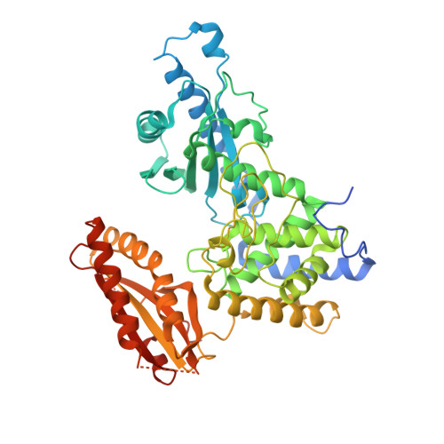
    4LT6

  • PubMed Abstract: 

    In eukaryotes, the poly(A) tail added at the 3' end of an mRNA precursor is essential for the regulation of mRNA stability and the initiation of translation. Poly(A) polymerase (PAP) is the enzyme that catalyzes the poly(A) addition reaction. Multiple isoforms of PAP have been identified in vertebrates, which originate from gene duplication, alternative splicing or post-translational modifications. The complexity of PAP isoforms suggests that they might play different roles in the cell. Phylogenetic studies indicate that vertebrate PAPs are grouped into three clades termed α, β and γ, which originated from two gene duplication events. To date, all the available PAP structures are from the PAPα clade. Here, we present the crystal structure of the first representative of the PAPγ clade, human PAPγ bound to cordycepin triphosphate (3'dATP) and Ca(2+). The structure revealed that PAPγ closely resembles its PAPα ortholog. An analysis of residue conservation reveals a conserved catalytic binding pocket, whereas residues at the surface of the polymerase are more divergent.


  • Organizational Affiliation

    Department of Microbiology and Molecular Genetics, University of Vermont, Stafford Hall, 95 Carrigan Drive, Burlington, VT 05405-0068, USA. Electronic address: yang@crystal.harvard.edu.


Macromolecules
Find similar proteins by:  (by identity cutoff)  |  3D Structure
Entity ID: 1
MoleculeChains Sequence LengthOrganismDetailsImage
Poly(A) polymerase gamma
A, B
514Homo sapiensMutation(s): 0 
Gene Names: PAPOLGPAP2PAPG
EC: 2.7.7.19
UniProt & NIH Common Fund Data Resources
Find proteins for Q9BWT3 (Homo sapiens)
Explore Q9BWT3 
Go to UniProtKB:  Q9BWT3
PHAROS:  Q9BWT3
GTEx:  ENSG00000115421 
Entity Groups  
Sequence Clusters30% Identity50% Identity70% Identity90% Identity95% Identity100% Identity
UniProt GroupQ9BWT3
Sequence Annotations
Expand
  • Reference Sequence
Experimental Data & Validation

Experimental Data

  • Method: X-RAY DIFFRACTION
  • Resolution: 2.79 Å
  • R-Value Free: 0.257 
  • R-Value Work: 0.193 
  • R-Value Observed: 0.196 
  • Space Group: P 21 21 21
Unit Cell:
Length ( Å )Angle ( ˚ )
a = 68.87α = 90
b = 89.92β = 90
c = 201.63γ = 90
Software Package:
Software NamePurpose
MAR345dtbdata collection
MOLREPphasing
PHENIXrefinement
DENZOdata reduction
SCALEPACKdata scaling

Structure Validation

View Full Validation Report


Ligand Structure Quality Assessment 


Entry History 

Deposition Data

Revision History  (Full details and data files)

  • Version 1.0: 2013-10-09
    Type: Initial release
  • Version 1.1: 2013-10-16
    Changes: Database references
  • Version 1.2: 2014-01-08
    Changes: Database references
  • Version 1.3: 2023-09-20
    Changes: Data collection, Database references, Derived calculations, Refinement description