4L1F

Electron transferring flavoprotein of Acidaminococcus fermentans: Towards a mechanism of flavin-based electron bifurcation


Experimental Data Snapshot

  • Method: X-RAY DIFFRACTION
  • Resolution: 1.79 Å
  • R-Value Free: 0.183 
  • R-Value Work: 0.153 
  • R-Value Observed: 0.155 

wwPDB Validation   3D Report Full Report


Ligand Structure Quality Assessment 


This is version 1.2 of the entry. See complete history


Literature

Studies on the Mechanism of Electron Bifurcation Catalyzed by Electron Transferring Flavoprotein (Etf) and Butyryl-CoA Dehydrogenase (Bcd) of Acidaminococcus fermentans.

Chowdhury, N.P.Mowafy, A.M.Demmer, J.K.Upadhyay, V.Koelzer, S.Jayamani, E.Kahnt, J.Hornung, M.Demmer, U.Ermler, U.Buckel, W.

(2014) J Biol Chem 289: 5145-5157

  • DOI: https://doi.org/10.1074/jbc.M113.521013
  • Primary Citation of Related Structures:  
    4KPU, 4L1F, 4L2I

  • PubMed Abstract: 

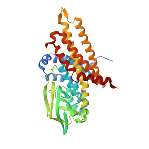
    Electron bifurcation is a fundamental strategy of energy coupling originally discovered in the Q-cycle of many organisms. Recently a flavin-based electron bifurcation has been detected in anaerobes, first in clostridia and later in acetogens and methanogens. It enables anaerobic bacteria and archaea to reduce the low-potential [4Fe-4S] clusters of ferredoxin, which increases the efficiency of the substrate level and electron transport phosphorylations. Here we characterize the bifurcating electron transferring flavoprotein (EtfAf) and butyryl-CoA dehydrogenase (BcdAf) of Acidaminococcus fermentans, which couple the exergonic reduction of crotonyl-CoA to butyryl-CoA to the endergonic reduction of ferredoxin both with NADH. EtfAf contains one FAD (α-FAD) in subunit α and a second FAD (β-FAD) in subunit β. The distance between the two isoalloxazine rings is 18 Å. The EtfAf-NAD(+) complex structure revealed β-FAD as acceptor of the hydride of NADH. The formed β-FADH(-) is considered as the bifurcating electron donor. As a result of a domain movement, α-FAD is able to approach β-FADH(-) by about 4 Å and to take up one electron yielding a stable anionic semiquinone, α-FAD, which donates this electron further to Dh-FAD of BcdAf after a second domain movement. The remaining non-stabilized neutral semiquinone, β-FADH(•), immediately reduces ferredoxin. Repetition of this process affords a second reduced ferredoxin and Dh-FADH(-) that converts crotonyl-CoA to butyryl-CoA.


  • Organizational Affiliation

    From the Laboratorium für Mikrobiologie, Fachbereich Biologie and SYNMIKRO, Philipps-Universität, 35032 Marburg, Germany.


Macromolecules
Find similar proteins by:  (by identity cutoff)  |  3D Structure
Entity ID: 1
MoleculeChains Sequence LengthOrganismDetailsImage
Acyl-CoA dehydrogenase domain protein
A, B
383Acidaminococcus fermentans DSM 20731Mutation(s): 0 
UniProt
Find proteins for D2RL84 (Acidaminococcus fermentans (strain ATCC 25085 / DSM 20731 / CCUG 9996 / CIP 106432 / VR4))
Explore D2RL84 
Go to UniProtKB:  D2RL84
Entity Groups  
Sequence Clusters30% Identity50% Identity70% Identity90% Identity95% Identity100% Identity
UniProt GroupD2RL84
Sequence Annotations
Expand
  • Reference Sequence
Small Molecules
Ligands 5 Unique
IDChains Name / Formula / InChI Key2D Diagram3D Interactions
COS
Query on COS

Download Ideal Coordinates CCD File 
D [auth A],
I [auth B]
COENZYME A PERSULFIDE
C21 H36 N7 O16 P3 S2
REVPHPVBPSIEKM-IBOSZNHHSA-N
FAD
Query on FAD

Download Ideal Coordinates CCD File 
C [auth A],
H [auth B]
FLAVIN-ADENINE DINUCLEOTIDE
C27 H33 N9 O15 P2
VWWQXMAJTJZDQX-UYBVJOGSSA-N
PO4
Query on PO4

Download Ideal Coordinates CCD File 
J [auth B]PHOSPHATE ION
O4 P
NBIIXXVUZAFLBC-UHFFFAOYSA-K
PDO
Query on PDO

Download Ideal Coordinates CCD File 
F [auth A]1,3-PROPANDIOL
C3 H8 O2
YPFDHNVEDLHUCE-UHFFFAOYSA-N
NA
Query on NA

Download Ideal Coordinates CCD File 
E [auth A],
G [auth A],
K [auth B]
SODIUM ION
Na
FKNQFGJONOIPTF-UHFFFAOYSA-N
Experimental Data & Validation

Experimental Data

  • Method: X-RAY DIFFRACTION
  • Resolution: 1.79 Å
  • R-Value Free: 0.183 
  • R-Value Work: 0.153 
  • R-Value Observed: 0.155 
  • Space Group: P 21 21 2
Unit Cell:
Length ( Å )Angle ( ˚ )
a = 109.1α = 90
b = 141.4β = 90
c = 64.1γ = 90
Software Package:
Software NamePurpose
HKL-2000data collection
SHARPphasing
REFMACrefinement
XDSdata reduction
XSCALEdata scaling

Structure Validation

View Full Validation Report


Ligand Structure Quality Assessment 


Entry History 

Deposition Data

Revision History  (Full details and data files)

  • Version 1.0: 2014-01-15
    Type: Initial release
  • Version 1.1: 2014-03-12
    Changes: Database references
  • Version 1.2: 2024-02-28
    Changes: Data collection, Database references, Derived calculations