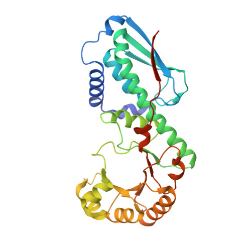
The crystal structure of human quinolinic acid phosphoribosyltransferase in complex with its inhibitor phthalic acid.
Malik, S.S., Patterson, D.N., Ncube, Z., Toth, E.A.(2014) Proteins 82: 405-414
- PubMed: 24038671
- DOI: https://doi.org/10.1002/prot.24406
- Primary Citation of Related Structures:
4KWV, 4KWW - PubMed Abstract:
Quinolinic acid (QA), a biologically potent but neurodestructive metabolite is catabolized by quinolinic acid phosphoribosyltransferase (QPRT) in the first step of the de novo NAD(+) biosynthesis pathway. This puts QPRT at the junction of two different pathways, that is, de novo NAD(+) biosynthesis and the kynurenine pathway of tryptophan degradation. Thus, QPRT is an important enzyme in terms of its biological impact and its potential as a therapeutic target. Here, we report the crystal structure of human QPRT bound to its inhibitor phthalic acid (PHT) and kinetic analysis of PHT inhibition of human QPRT. This structure, determined at 2.55 Å resolution, shows an elaborate hydrogen bonding network that helps in recognition of PHT and consequently its substrate QA. In addition to this hydrogen bonding network, we observe extensive van der Waals contacts with the PHT ring that might be important for correctly orientating the substrate QA during catalysis. Moreover, our crystal form allows us to observe an intact hexamer in both the apo- and PHT-bound forms in the same crystal system, which provides a direct comparison of unique subunit interfaces formed in hexameric human QPRT. We call these interfaces "nondimeric interfaces" to distinguish them from the typical dimeric interfaces observed in all QPRTs. We observe significant changes in the nondimeric interfaces in the QPRT hexamer upon binding PHT. Thus, the new structural and functional features of this enzyme we describe here will aid in understanding the function of hexameric QPRTs, which includes all eukaryotic and select prokaryotic QPRTs.
Organizational Affiliation:
Department of Biochemistry and Molecular Biology, University of Maryland School of Medicine, Baltimore, Maryland 21201.