4KFV

Structural insight into Golgi membrane stacking by GRASP65 and GRASP55


Experimental Data Snapshot

  • Method: X-RAY DIFFRACTION
  • Resolution: 2.20 Å
  • R-Value Free: 0.283 
  • R-Value Work: 0.214 
  • R-Value Observed: 0.217 

wwPDB Validation   3D Report Full Report


This is version 1.3 of the entry. See complete history


Literature

Structural insight into Golgi membrane stacking by GRASP65 and GRASP55 proteins

Feng, Y.Yu, W.Li, X.Lin, S.Zhou, Y.Hu, J.Liu, X.

(2013) J Biol Chem 288: 28418-28427

  • DOI: https://doi.org/10.1074/jbc.M113.478024
  • Primary Citation of Related Structures:  
    4KFV, 4KFW

  • PubMed Abstract: 

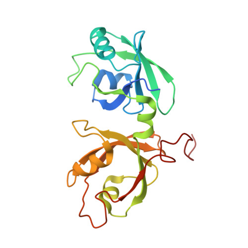
    The stacking of Golgi cisternae involves GRASP65 and GRASP55. The oligomerization of the N-terminal GRASP domain of these proteins, which consists of two tandem PDZ domains, is required to tether the Golgi membranes. However, the molecular basis for GRASP assembly is unclear. Here, we determined the crystal structures of the GRASP domain of GRASP65 and GRASP55. The structures reveal similar homotypic interactions: the GRASP domain forms a dimer in which the peptide-binding pockets of the two neighboring PDZ2 domains face each other, and the dimers are further connected by the C-terminal tail of one GRASP domain inserting into the binding pocket of the PDZ1 domain in another dimer. Biochemical analysis suggests that both types of contacts are relatively weak but are needed in combination for GRASP-mediated Golgi stacking. Our results unveil a novel mode of membrane tethering by GRASP proteins and provide insight into the mechanism of Golgi stacking.


  • Organizational Affiliation

    From the Department of Biochemistry and Molecular Biology, College of Life Sciences, and State Key Laboratory of Medicinal Chemical Biology and.


Macromolecules
Find similar proteins by:  (by identity cutoff)  |  3D Structure
Entity ID: 1
MoleculeChains Sequence LengthOrganismDetailsImage
Golgi reassembly-stacking protein 1210Rattus norvegicusMutation(s): 0 
Gene Names: Gorasp1GRASP 65rCG_25352
UniProt
Find proteins for O35254 (Rattus norvegicus)
Explore O35254 
Go to UniProtKB:  O35254
Entity Groups  
Sequence Clusters30% Identity50% Identity70% Identity90% Identity95% Identity100% Identity
UniProt GroupO35254
Sequence Annotations
Expand
  • Reference Sequence
Experimental Data & Validation

Experimental Data

  • Method: X-RAY DIFFRACTION
  • Resolution: 2.20 Å
  • R-Value Free: 0.283 
  • R-Value Work: 0.214 
  • R-Value Observed: 0.217 
  • Space Group: P 21 21 2
Unit Cell:
Length ( Å )Angle ( ˚ )
a = 44.989α = 90
b = 104.287β = 90
c = 37.932γ = 90
Software Package:
Software NamePurpose
HKL-2000data collection
PHENIXrefinement
HKL-2000data reduction
HKL-2000data scaling

Structure Validation

View Full Validation Report


Entry History 

Deposition Data

  • Released Date: 2013-08-21 
  • Deposition Author(s): Liu, X., Hu, J.

Revision History  (Full details and data files)

  • Version 1.0: 2013-08-21
    Type: Initial release
  • Version 1.1: 2014-01-22
    Changes: Database references
  • Version 1.2: 2017-11-15
    Changes: Refinement description
  • Version 1.3: 2024-03-20
    Changes: Data collection, Database references, Derived calculations